Copyright
©2014 Baishideng Publishing Group Inc.
World J Cardiol. Sep 26, 2014; 6(9): 959-967
Published online Sep 26, 2014. doi: 10.4330/wjc.v6.i9.959
Published online Sep 26, 2014. doi: 10.4330/wjc.v6.i9.959
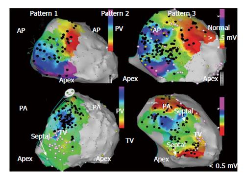
Figure 1 Bipolar right ventricle endocardial voltage maps demonstrating characteristic patterns of low voltage (< 1.
5 mV) regions identified in patients with arrhythmogenic right ventricular cardiomyopathy/dysplasi and ventricular tachycardia in anterior and posterior views. Peritricuspid (pattern 1), peripulmonic (pattern 2), or more extensive involvement extending from both valvular regions (pattern 3) is shown. Distribution of abnormal electrograms is predominantly free wall. Right ventricle apex is spared, and septal involvement is frequently identified (arrows). Adapted from Marchlinski et al[15] with permission. AP: Anterior; PA: Posterior.
- Citation: Tschabrunn CM, Marchlinski FE. Ventricular tachycardia mapping and ablation in arrhythmogenic right ventricular cardiomyopathy/dysplasia: Lessons Learned. World J Cardiol 2014; 6(9): 959-967
- URL: https://www.wjgnet.com/1949-8462/full/v6/i9/959.htm
- DOI: https://dx.doi.org/10.4330/wjc.v6.i9.959


