Copyright
©2014 Baishideng Publishing Group Inc.
World J Cardiol. Jul 26, 2014; 6(7): 585-601
Published online Jul 26, 2014. doi: 10.4330/wjc.v6.i7.585
Published online Jul 26, 2014. doi: 10.4330/wjc.v6.i7.585
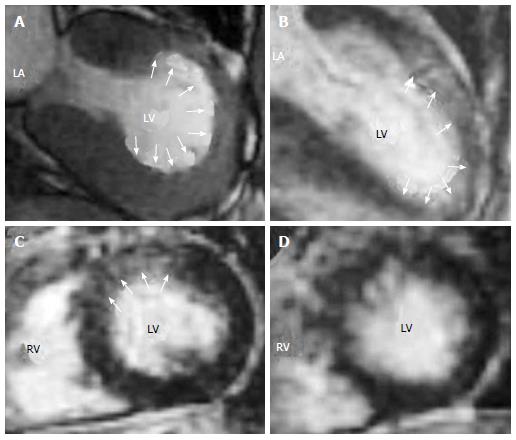
Figure 6 Representative cine-cardiac magnetic resonance (A) and late gadolinium enhancement-cardiac magnetic resonance (B-D) images in a case of stress (Takotsubo) cardiomyopathy.
The images show vertical long axis (A, B) and mid-ventricular short axis (C, D) views. The cine-CMR image during systole (A) shows mid-anterior dyskinesis (white arrows). LGE-CMR images on the sub-acute phase (B, C) show that the area of LGE was well matched with the area of wall motion abnormality (white arrows). On the follow-up phase, LV systolic function recovered, and the LGE-CMR image (D) could not detect significant LGE in the LGE area observed on the sub-acute phase. All the images are taken from Naruse et al[69] with permission. LGE: Late gadolinium enhancement; LV: Left ventricular; LGE-CMR: Late gadolinium enhancement-cardiac magnetic resonance.
- Citation: Satoh H, Sano M, Suwa K, Saitoh T, Nobuhara M, Saotome M, Urushida T, Katoh H, Hayashi H. Distribution of late gadolinium enhancement in various types of cardiomyopathies: Significance in differential diagnosis, clinical features and prognosis. World J Cardiol 2014; 6(7): 585-601
- URL: https://www.wjgnet.com/1949-8462/full/v6/i7/585.htm
- DOI: https://dx.doi.org/10.4330/wjc.v6.i7.585


