Copyright
©2014 Baishideng Publishing Group Inc.
World J Cardiol. Jul 26, 2014; 6(7): 585-601
Published online Jul 26, 2014. doi: 10.4330/wjc.v6.i7.585
Published online Jul 26, 2014. doi: 10.4330/wjc.v6.i7.585
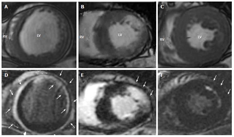
Figure 4 Representative cine-cardiac magnetic resonance (A-C) and late gadolinium enhancement-cardiac magnetic resonance (D-F) images in patients with cardiac sarcoidosis.
A, D: A patient with LV dilatation, reduced LVEF (22%) and circumferential subepicardial and subendocardial LGE with spared mid-myocardium; B, E: A patient with reduced LVEF (38%) and nodular LGE in antero-lateral wall; C, F: A patient with preserved LVEF (58%) with mid-wall striated LGE in antero-lateral wall. White arrows indicate LGE areas. A part of the images is taken from Matoh et al[10] with permission. LGE: Late gadolinium enhancement; LV: Left ventricular; LVEF: Left ventricular ejection fraction
- Citation: Satoh H, Sano M, Suwa K, Saitoh T, Nobuhara M, Saotome M, Urushida T, Katoh H, Hayashi H. Distribution of late gadolinium enhancement in various types of cardiomyopathies: Significance in differential diagnosis, clinical features and prognosis. World J Cardiol 2014; 6(7): 585-601
- URL: https://www.wjgnet.com/1949-8462/full/v6/i7/585.htm
- DOI: https://dx.doi.org/10.4330/wjc.v6.i7.585


