Copyright
©2014 Baishideng Publishing Group Inc.
World J Cardiol. Jul 26, 2014; 6(7): 585-601
Published online Jul 26, 2014. doi: 10.4330/wjc.v6.i7.585
Published online Jul 26, 2014. doi: 10.4330/wjc.v6.i7.585
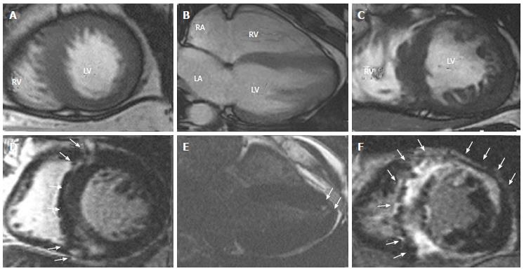
Figure 3 Representative cine-cardiac magnetic resonance (A-C) and late gadolinium enhancement-cardiac magnetic resonance (D-F) images in patients with various phenotypes of hypertrophic cardiomyopathy.
A, D: ASH (short axis views); B, E: APH (horizontal views); C, F: End-stage HCM (short axis views). LGE was mainly localized in the ventricular septum and right ventricular insertion points in ASH and in the apex in APH (arrows). Note the inhomogeneous LV wall thickness and diffusely spread LGE in end-stage HCM. All the images are taken from Satoh et al[11]. ASH: Asymmetrical septal hypertrophy; APH: Apical hypertrophy; LGE: Late gadolinium enhancement; LV: Left ventricular; HCM: Hypertrophic cardiomyopathy.
- Citation: Satoh H, Sano M, Suwa K, Saitoh T, Nobuhara M, Saotome M, Urushida T, Katoh H, Hayashi H. Distribution of late gadolinium enhancement in various types of cardiomyopathies: Significance in differential diagnosis, clinical features and prognosis. World J Cardiol 2014; 6(7): 585-601
- URL: https://www.wjgnet.com/1949-8462/full/v6/i7/585.htm
- DOI: https://dx.doi.org/10.4330/wjc.v6.i7.585


