Copyright
©2014 Baishideng Publishing Group Inc.
World J Cardiol. Jul 26, 2014; 6(7): 585-601
Published online Jul 26, 2014. doi: 10.4330/wjc.v6.i7.585
Published online Jul 26, 2014. doi: 10.4330/wjc.v6.i7.585
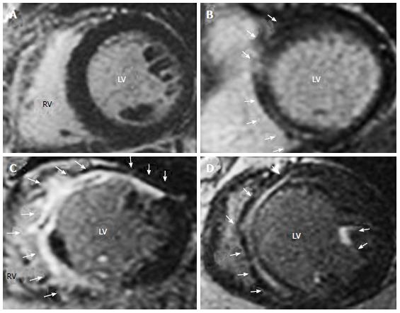
Figure 2 Representative short axis late gadolinium enhancement-cardiac magnetic resonance images in patients with dilated cardiomyopathy.
A: No LGE; B: localized LGE. Mid-wall LGE distributed only into anterior and inferior septum; C: Extensive LGE. LGE distributed at anterior and inferior septum, anterior, antero-lateral and inferior LV segments; D: Extensive LGE. Mid wall LGE distributed at anterior and inferior septum, and at anterior papillary muscle. Arrows indicate LGE in LV wall segments. All the images are taken from Machii et al[13] with permission. LGE: Late gadolinium enhancement; LV: Left ventricular.
- Citation: Satoh H, Sano M, Suwa K, Saitoh T, Nobuhara M, Saotome M, Urushida T, Katoh H, Hayashi H. Distribution of late gadolinium enhancement in various types of cardiomyopathies: Significance in differential diagnosis, clinical features and prognosis. World J Cardiol 2014; 6(7): 585-601
- URL: https://www.wjgnet.com/1949-8462/full/v6/i7/585.htm
- DOI: https://dx.doi.org/10.4330/wjc.v6.i7.585


