Copyright
©2014 Baishideng Publishing Group Inc.
World J Cardiol. Jul 26, 2014; 6(7): 577-584
Published online Jul 26, 2014. doi: 10.4330/wjc.v6.i7.577
Published online Jul 26, 2014. doi: 10.4330/wjc.v6.i7.577
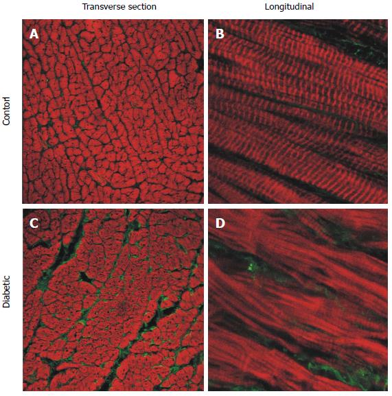
Figure 5 Representative confocal images of longitudinal section free wall immuno-labelled for type I collagen (green) and f-actin (red).
Sections from the endocardium of control (A and B) and diabetic (C and D) rat hearts. Left hand side panels: Transverse sections from endocardium (25 × objective). Right hand side panels: Longitudinal sections (63 × objective, zoom × 3). (Modified from Zhang et al[23]).
- Citation: Ward ML, Crossman DJ. Mechanisms underlying the impaired contractility of diabetic cardiomyopathy. World J Cardiol 2014; 6(7): 577-584
- URL: https://www.wjgnet.com/1949-8462/full/v6/i7/577.htm
- DOI: https://dx.doi.org/10.4330/wjc.v6.i7.577


