Copyright
©2014 Baishideng Publishing Group Co.
World J Cardiol. Apr 26, 2014; 6(4): 183-195
Published online Apr 26, 2014. doi: 10.4330/wjc.v6.i4.183
Published online Apr 26, 2014. doi: 10.4330/wjc.v6.i4.183
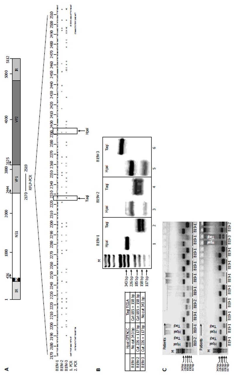
Figure 1 Generation of B19V-genotype 1 to 3 specific restriction fragment length polymorphism-polymerase chain reaction.
A: Schematic representation of the B19V genome showing localization of the B19V-genotype-specific restriction fragment length polymorphism-polymerase chain reaction (RFLP-PCR) in the B19V NS1-VP1u region. Sequences of B19V-1, B19V-2 and B19V-3 showing the RFLP-PCR fragment and HpaI and TaqI restriction enzyme sites (lower panel). Primer positions for 1st and 2nd RFLP-PCR are indicated (1. PCR and 2. PCR, see also Table 2); B: Expected fragment size and digestion pattern after HpaI and TaqI digestions (right panel). Agarose gel electrophoresis showing respective PCR-fragments after HpaI and TaqI digestion for each B19V-genotype (left panel); C: Representative agarose gel electrophoresis of patient-specific B19V RFLP-PCRs. B19V-1: B19V-genotype 1.
- Citation: Bock CT, Düchting A, Utta F, Brunner E, Sy BT, Klingel K, Lang F, Gawaz M, Felix SB, Kandolf R. Molecular phenotypes of human parvovirus B19 in patients with myocarditis. World J Cardiol 2014; 6(4): 183-195
- URL: https://www.wjgnet.com/1949-8462/full/v6/i4/183.htm
- DOI: https://dx.doi.org/10.4330/wjc.v6.i4.183


