Copyright
©The Author(s) 2023.
World J Cardiol. Sep 26, 2023; 15(9): 415-426
Published online Sep 26, 2023. doi: 10.4330/wjc.v15.i9.415
Published online Sep 26, 2023. doi: 10.4330/wjc.v15.i9.415
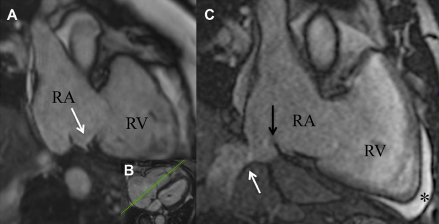
Figure 6 Isthmus pouches that are frequently present, a prominent Eustachian ridge and large pectinate muscles may impede catheter stability and navigation to target sites leading to poor tissue contact and low radio frequency energy delivery.
A and B: Septal pouch (white arrow) of the cavotricuspid isthmus in a balanced steady-state free precision sequence image [as visualized in the transversal plane (B)]; C: Kinking of the vena cava inferior junction in the right atrium (white arrow) and a eustachian valve (black arrow) was found. Anterior to the right ventricular apex, a pre-existing pericardial effusion was located (asterisk). Citation: Ulbrich S, Huo Y, Tomala J, Wagner M, Richter U, Pu L, Mayer J, Zedda A, Krafft AJ, Lindborg K, Piorkowski C, Gaspar T. Magnetic resonance imaging-guided conventional catheter ablation of isthmus-dependent atrial flutter using active catheter imaging. Heart Rhythm O2 2022; 3: 553-559 [PMID: 36340492 DOI: 10.1016/j.hroo.2022.06.011]. Copyright © 2022 Heart Rhythm Society. Published by Elsevier Inc. (Reproduced under the terms of the Creative Commons CC-BY license)[40].
- Citation: Tampakis K, Pastromas S, Sykiotis A, Kampanarou S, Kourgiannidis G, Pyrpiri C, Bousoula M, Rozakis D, Andrikopoulos G. Real-time cardiovascular magnetic resonance-guided radiofrequency ablation: A comprehensive review. World J Cardiol 2023; 15(9): 415-426
- URL: https://www.wjgnet.com/1949-8462/full/v15/i9/415.htm
- DOI: https://dx.doi.org/10.4330/wjc.v15.i9.415


