Copyright
©The Author(s) 2022.
World J Cardiol. Apr 26, 2022; 14(4): 190-205
Published online Apr 26, 2022. doi: 10.4330/wjc.v14.i4.190
Published online Apr 26, 2022. doi: 10.4330/wjc.v14.i4.190
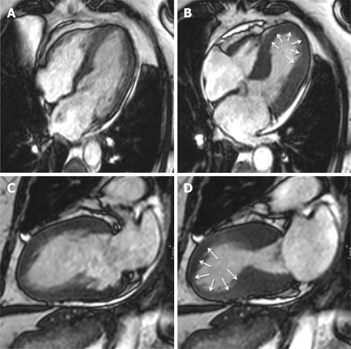
Figure 2 Cardiac magnetic resonance imaging of Takotsubo cardiomyopathy.
Typical apical ballooning seen in takotsubo syndrome. A, B: Cine four-chamber in late diastole and systole respectively; C, D: Two-chamber view in late diastole and systole respectively. Modified from Plácido et al[123] and licensed under the Creative Commons Attribution 4.0 International License (http://creativecommons.org/licenses/by/4.0/).
- Citation: Nguyen Nguyen N, Assad JG, Femia G, Schuster A, Otton J, Nguyen TL. Role of cardiac magnetic resonance imaging in troponinemia syndromes. World J Cardiol 2022; 14(4): 190-205
- URL: https://www.wjgnet.com/1949-8462/full/v14/i4/190.htm
- DOI: https://dx.doi.org/10.4330/wjc.v14.i4.190


