Copyright
©The Author(s) 2018.
World J Cardiol. Oct 26, 2018; 10(10): 165-186
Published online Oct 26, 2018. doi: 10.4330/wjc.v10.i10.165
Published online Oct 26, 2018. doi: 10.4330/wjc.v10.i10.165
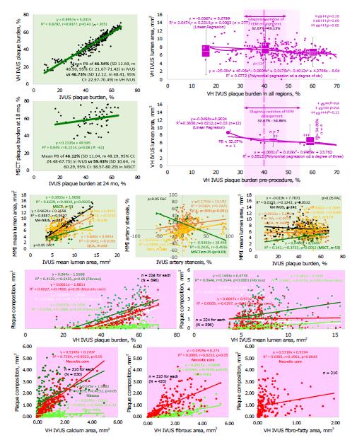
Figure 6 The association between the components of the lesion and artery layers.
The correlation between plaque burden (PB) assessed by intravascular ultrasound (IVUS) and virtual histology (VH)-IVUS was strong with relatively weak association with PB evaluated by multislice computed tomography (MSCT) (top left panel). The three-distribution (PB < 32.07%, PB 32.07%-49.13%, and PB > 49.13%) box-and-whisker analysis (top right panel separately for all regions and pre-procedure) of the VH-IVUS-examined correlation between PB and lumen area vindicated existence of the window of the external elastic membrane (EEM) enlargement between 32.07% and 49.13%. The pre-procedure evaluation in naïve arteries with a broader size of the window between 32.07% and 54.86%. The middle and bottom panels set out correlations between plaque burden, lumen, vessel wall dimensions, and components of the lesion examined by the various imaging modalities. The P value was calculated for comparison of one or two variables in order to either estimate the means of two groups (paired or unpaired t test) or appreciate statistical consistency for regression. The figure was adapted from ref. [34]. n: Number of observations; N: Total number of observations at the screened population; R2: Coefficient of determination; r: Pearson correlation (for linear regression); IQR: Interquartile range; NS: Non-significant (P > 0.05); NA: Not applicable.
- Citation: Kharlamov AN. Undiscovered pathology of transient scaffolding t1remains a driver of failures in clinical trials. World J Cardiol 2018; 10(10): 165-186
- URL: https://www.wjgnet.com/1949-8462/full/v10/i10/165.htm
- DOI: https://dx.doi.org/10.4330/wjc.v10.i10.165


