Copyright
©The Author(s) 2017.
World J Biol Chem. Feb 26, 2017; 8(1): 57-80
Published online Feb 26, 2017. doi: 10.4331/wjbc.v8.i1.57
Published online Feb 26, 2017. doi: 10.4331/wjbc.v8.i1.57
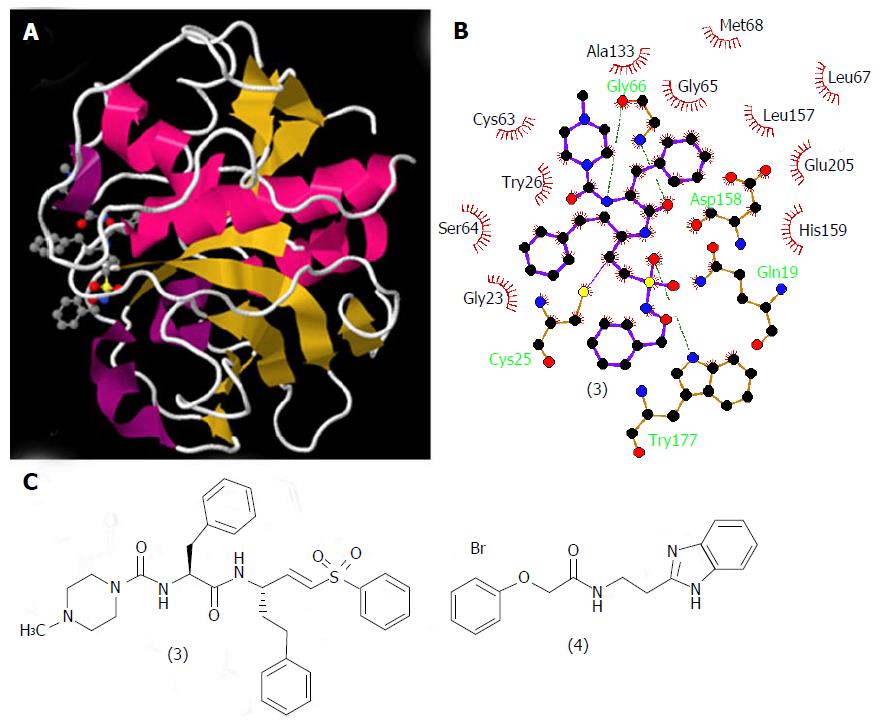
Figure 5 Structures of cruzain and its ligands.
A: Three-dimensional structural representation of cruzain crystallized with vinyl sulfone derivative (3). Structure deposited in the Protein Data Bank under the code 1F2C[45]; B: Schematic representation in two dimensions of the binding mode of (3) to cruzain generated by LigPlot+ software upon the PDB file; C: Structures of the cruzain inhibitor derivatives (3) and (4).
- Citation: Sueth-Santiago V, Decote-Ricardo D, Morrot A, Freire-de-Lima CG, Lima MEF. Challenges in the chemotherapy of Chagas disease: Looking for possibilities related to the differences and similarities between the parasite and host. World J Biol Chem 2017; 8(1): 57-80
- URL: https://www.wjgnet.com/1949-8454/full/v8/i1/57.htm
- DOI: https://dx.doi.org/10.4331/wjbc.v8.i1.57


