Copyright
©The Author(s) 2017.
World J Biol Chem. Feb 26, 2017; 8(1): 32-44
Published online Feb 26, 2017. doi: 10.4331/wjbc.v8.i1.32
Published online Feb 26, 2017. doi: 10.4331/wjbc.v8.i1.32
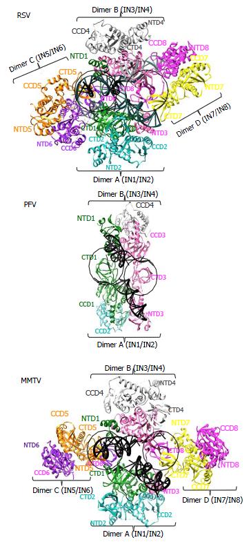
Figure 4 Integrase domain organizations within the prototype foamy virus, Rous sarcoma virus and mouse mammary tumor virus intasome structures.
Separate integrase (IN) domains are labeled, with IN monomer coloring code retained from Figure 3. The green IN1 and pink IN3 monomers donate their active sites for catalysis of 3’ processing and strand transfer across the structures. Circled areas represent similarly positioned CTDs. While these emanate from inner IN1 and IN3 monomers in the PFV structure, they originate from flanking MMTV and RSV IN monomers IN6 and IN8. PDB accession codes same as in Figure 3. PFV: Prototype foamy virus; RSV: Rous sarcoma virus; MMTV: Mouse mammary tumor virus; CCD: Catalytic core domain; NTD: N-terminal domain; CTD: C-terminal domain.
- Citation: Grawenhoff J, Engelman AN. Retroviral integrase protein and intasome nucleoprotein complex structures. World J Biol Chem 2017; 8(1): 32-44
- URL: https://www.wjgnet.com/1949-8454/full/v8/i1/32.htm
- DOI: https://dx.doi.org/10.4331/wjbc.v8.i1.32


