Copyright
©2014 Baishideng Publishing Group Inc.
World J Biol Chem. Aug 26, 2014; 5(3): 334-345
Published online Aug 26, 2014. doi: 10.4331/wjbc.v5.i3.334
Published online Aug 26, 2014. doi: 10.4331/wjbc.v5.i3.334
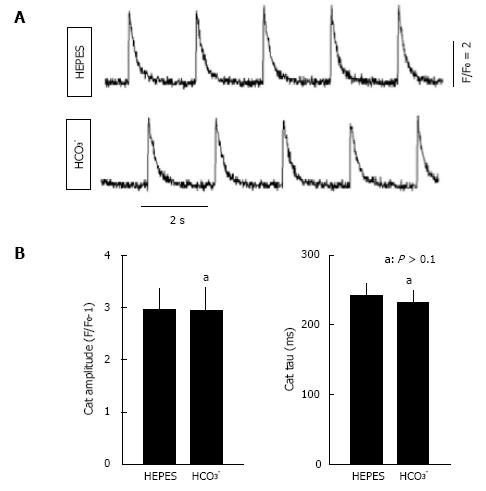
Figure 3 Ca2+ transient analysis in isolated rat myocytes bathed in CO2/HCO3- buffer and in HEPES buffer.
For recording of Ca2+ transients, isolated ventricular myocytes were loaded with fluo-4 acetoxymethyl ester (5 μmol/L, Molecular Probes, Eugene, OR) and activated with field stimulation at 0.5 Hz. Fluorescence signals were measured using a Nikon TE 2000 microscope and an InCyt Standard PM photometry system (Intracellular Imaging, Cincinnati, OH) as described previously[104]. A: Representative Ca2+ transients in HEPES and HCO3--containing buffers; B: Average Ca2+ transient (CaT) amplitudes and tau values, a measure of the rate of decay of the Ca2+ transient in HEPES and HCO3--containing buffers (n = 12). The same cells were imaged in both buffers. Myocytes were from the same preparations used in Figure 2, with statistical analyses performed using a paired t-test. No significant differences were observed.
- Citation: Wang HS, Chen Y, Vairamani K, Shull GE. Critical role of bicarbonate and bicarbonate transporters in cardiac function. World J Biol Chem 2014; 5(3): 334-345
- URL: https://www.wjgnet.com/1949-8454/full/v5/i3/334.htm
- DOI: https://dx.doi.org/10.4331/wjbc.v5.i3.334


