Copyright
©2014 Baishideng Publishing Group Inc.
World J Biol Chem. May 26, 2014; 5(2): 204-215
Published online May 26, 2014. doi: 10.4331/wjbc.v5.i2.204
Published online May 26, 2014. doi: 10.4331/wjbc.v5.i2.204
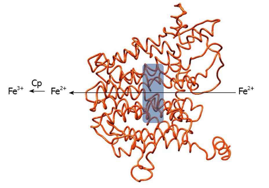
Figure 1 Structural model of human ferroportin viewed along the membrane plane.
The gray box indicates the location of a putative iron-binding site, ferrous iron flows through the protein from the cell interior and is then oxidized by ceruloplasmin at the extracellular side. The figure was produced with Chimera[96].
- Citation: Musci G, Polticelli F, Bonaccorsi di Patti MC. Ceruloplasmin-ferroportin system of iron traffic in vertebrates. World J Biol Chem 2014; 5(2): 204-215
- URL: https://www.wjgnet.com/1949-8454/full/v5/i2/204.htm
- DOI: https://dx.doi.org/10.4331/wjbc.v5.i2.204


