Copyright
©2012 Baishideng Publishing Group Co.
World J Biol Chem. Jan 26, 2012; 3(1): 7-26
Published online Jan 26, 2012. doi: 10.4331/wjbc.v3.i1.7
Published online Jan 26, 2012. doi: 10.4331/wjbc.v3.i1.7
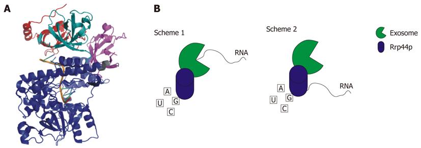
Figure 4 Rrp44p.
A: Crystal structure of Saccharomyces cerevisiae Rrp44p bound to RNA. CSD1 is colored red, CSD2 is colored teal, the RNB domain is blue, and the S1 domain is magenta. The PIN domain is not pictured because this domain is not included in the crystallized construct (RCSB # 2VNU); B: Schematic representation of the two mechanisms whereby Rrp44p is able to degrade RNA. In Scheme 1 the RNA is fed through the exosome to Rrp44p. Scheme 2 shows the RNA being degraded directly by Rrp44p[4,144].
- Citation: Bernstein J, Toth EA. Yeast nuclear RNA processing. World J Biol Chem 2012; 3(1): 7-26
- URL: https://www.wjgnet.com/1949-8454/full/v3/i1/7.htm
- DOI: https://dx.doi.org/10.4331/wjbc.v3.i1.7


