Copyright
©2010 Baishideng Publishing Group Co.
World J Biol Chem. May 26, 2010; 1(5): 144-150
Published online May 26, 2010. doi: 10.4331/wjbc.v1.i5.144
Published online May 26, 2010. doi: 10.4331/wjbc.v1.i5.144
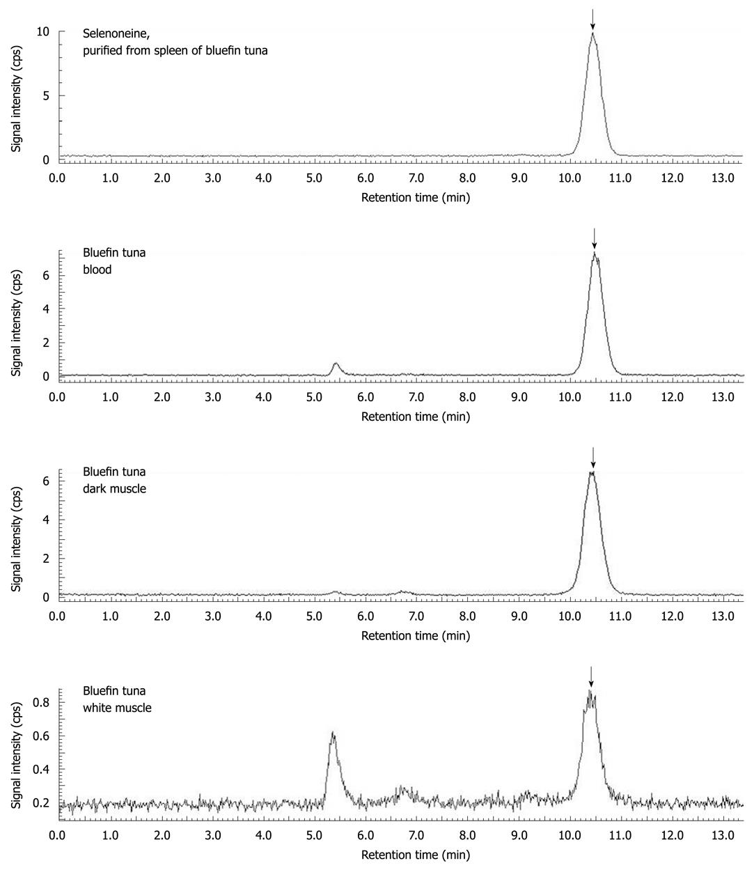
Figure 3 Speciation analysis of organic selenium in the various tissues of bluefin tuna by LC-ICP-MS.
Selenium compounds were separated with an Ultrahydrogel 120 (7.8 mm × 250 mm, Nihon Waters, Tokyo, Japan), equilibrated with 100 mmol/L ammonium formate buffer[30]. The mobile phase was delivered at 1 mL/min isocratically, and selenium was detected using online LC-ICP-MS (ELAN DRC II, PerkinElmer, Waltham, MA, USA) monitoring 82Se. GPx, selenite, selenocysteine, selenomethionine, and selenoneine were eluted at retention times of 5.4, 7.4, 7.8, 9.8, and 10.5 min, respectively, and the selenium concentration was determined using the respective compounds as standards. The arrow shows the elution of selenoneine. Selenoproteins including GPx were eluted near the void volume of the column at 5.4 min.
- Citation: Yamashita Y, Yabu T, Yamashita M. Discovery of the strong antioxidant selenoneine in tuna and selenium redox metabolism. World J Biol Chem 2010; 1(5): 144-150
- URL: https://www.wjgnet.com/1949-8454/full/v1/i5/144.htm
- DOI: https://dx.doi.org/10.4331/wjbc.v1.i5.144


