Copyright
©2010 Baishideng Publishing Group Co.
World J Biol Chem. May 26, 2010; 1(5): 133-143
Published online May 26, 2010. doi: 10.4331/wjbc.v1.i5.133
Published online May 26, 2010. doi: 10.4331/wjbc.v1.i5.133
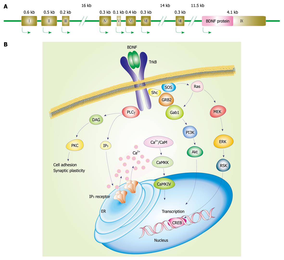
Figure 1 Brain-derived neurotrophic factor (BDNF) gene and stimulated intracellular signaling cascades after activation of tropomyosin-related kinase (Trk)B.
A: Mouse and rat BDNF genes (we referred to the description by Aid et al[15]). Each BDNF transcript is comprised of one of eight 5’ untranslated exons (exon I-VIII) and the common 3’ protein coding exon IX; B: Intracellular signaling after TrkB activation. Following BDNF binding, TrkB dimerization and its phosphorylation at intracellular tyrosine residues occur. Then, the activated TrkB stimulates three main signaling pathways: (1) mitogen-activated protein kinase/extracellular signal-regulated kinase (MAPK/ERK); (2) phosphatidylinositol 3-kinase (PI3K); and (3) phospholipase Cγ (PLCγ) pathways. MAPK pathway, in which MAPK/ERK kinase (MEK) is involved, plays a role in the neuronal differentiation and outgrowth. PI3K signaling promotes neuronal survival via Ras or GRB-associated binder 1 (Gab1). Following PLCγ activation, inositol-1,4,5-trisphosphate (IP3) and diacylglycerol (DAG) are both produced. DAG activates protein kinase C (PKC), which is important for regulation of synaptic plasticity. Meanwhile, IP3 increases intracellular Ca2+ concentration via IP3 receptors on the endoplasmic reticulum (ER), resulting in activation of Ca2+/calmodulin (CaM)-dependent protein kinase including CaMKII, CaMKK, and CaMKI. These MAPK/ERK, PI3K, and PLCγ pathways can regulate gene transcription.
- Citation: Numakawa T, Yokomaku D, Richards M, Hori H, Adachi N, Kunugi H. Functional interactions between steroid hormones and neurotrophin BDNF. World J Biol Chem 2010; 1(5): 133-143
- URL: https://www.wjgnet.com/1949-8454/full/v1/i5/133.htm
- DOI: https://dx.doi.org/10.4331/wjbc.v1.i5.133


