Copyright
©2010 Baishideng Publishing Group Co.
World J Biol Chem. May 26, 2010; 1(5): 109-132
Published online May 26, 2010. doi: 10.4331/wjbc.v1.i5.109
Published online May 26, 2010. doi: 10.4331/wjbc.v1.i5.109
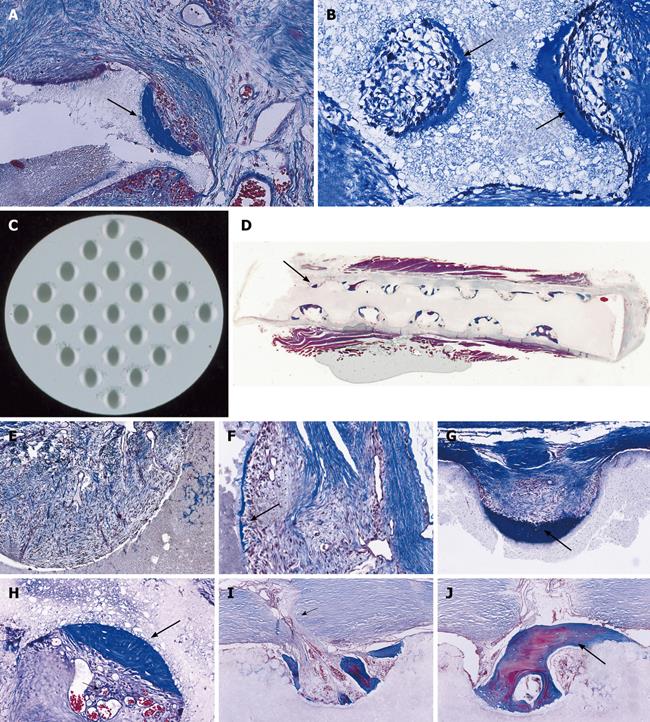
Figure 10 Influence of geometry on bone induction and morphogenesis: self inducing geometric cues initiating the induction of bone without the exogenous application of the osteogenic proteins of the TGF-β supergene family.
A, B: Macroporous sintered crystalline hydroxyapatites and the spontaneous induction of bone formation within concavities (thick long arrows) of the heterotopically implanted substratum 30 d after implantation. Based on the digital images of Figure 9E, G and of the above digital microphotographs, solid discs of highly crystalline hydroxyapatites with a series of concavities on both planar surfaces (C) were constructed and implanted in the rectus abdominis muscle of adult non-human primates of the species P. ursinus; D: Histological analyses of the harvested discs show the reproducible induction of bone formation within the concavities of the substratum only (thick long arrow); bone initiates only within the pre-cut concavities of the biomimetic matrix; the images were responsible for the definition of the phenomenon of the “geometric induction of bone formation”[82,83]; E, F: Middle power views of vascular and mesenchymal tissue invasion on day 30 within concavities of highly crystalline sintered hydroxyapatites with capillary sprouting and the beginning of the induction of bone formation (arrow in F) attached to the implanted substratum; G: Thick long arrow point to newly induced bone on day 90 after implantation of the biomimetic matrix; H-J: Remodeling, growth and pattern formation of the newly formed bone within concavities of highly crystalline sintered hydroxyapatites with prominent vascular invasion (short arrows in H and I) feeding the newly formed and remodeled bone initiated within the concavities of the implanted substrata (thick long arrows in H and J). Decalcified sections cut at 6 μm and stained with Goldner’s trichrome.
- Citation: Ripamonti U. Soluble and insoluble signals sculpt osteogenesis in angiogenesis. World J Biol Chem 2010; 1(5): 109-132
- URL: https://www.wjgnet.com/1949-8454/full/v1/i5/109.htm
- DOI: https://dx.doi.org/10.4331/wjbc.v1.i5.109


