Copyright
©2010 Baishideng Publishing Group Co.
World J Biol Chem. May 26, 2010; 1(5): 109-132
Published online May 26, 2010. doi: 10.4331/wjbc.v1.i5.109
Published online May 26, 2010. doi: 10.4331/wjbc.v1.i5.109
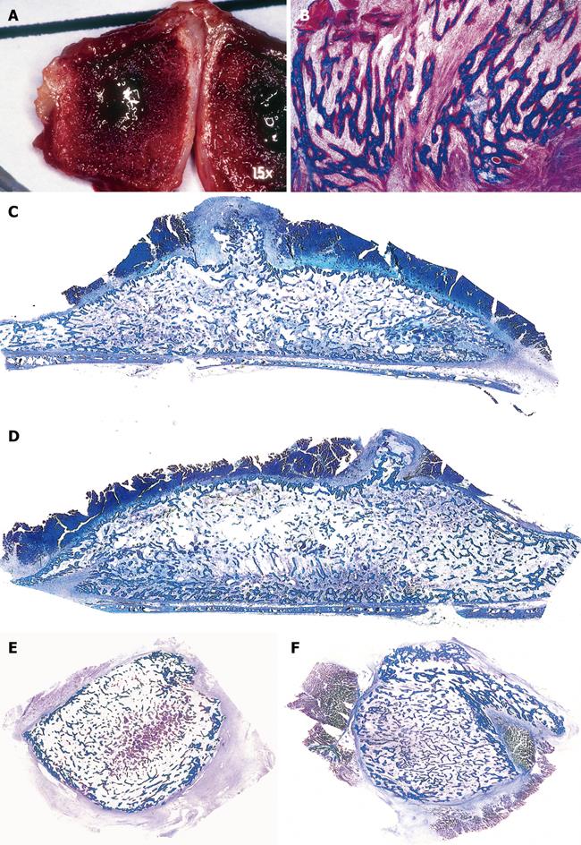
Figure 7 Prominent induction of bone formation by binary applications of recombinant hOP-1 with relatively low doses of recombinant hTGF-β1 implanted in the rectus abdominis muscle of P.
ursinus. A, B: Morphology of tissue induction and rapid tissue growth by binary application of 25 μg hOP-1 and 0.5 μg hTGF-β1 harvested on day 15 after implantation in the rectus abdominis muscle. A: Induction of a corticalized ossicle on day 15 after the synergistic binary implantation in the rectus abdominis muscle; B: Trabeculae of mineralized bone in blue on day 15 are surfaced by osteoid seams (red-orange) populated by contiguous osteoblasts; C, D: Prominent induction of heterotopic bone after implantation of 25 μg hOP-1 contra laterally juxtaposed to doses of 5 μg[68] across the rectus abdominis muscle of adult non-human primates of the species P. ursinus[68]. The radius of activity of the singly implanted hTGF-β1 osteogenic devices synergistically enhanced the inductive activity of the contra laterally implanted 25 μg hOP-1 devices[68]; E, F: Large heterotopic mineralized and corticalized ossicles generated by binary application of 25 μg hOP-1 and 0.5 μg hTGF-β1 and harvested on day 30. Undecalcified sections cut at 6 μm stained free-floating with a modified Goldner’s trichrome.
- Citation: Ripamonti U. Soluble and insoluble signals sculpt osteogenesis in angiogenesis. World J Biol Chem 2010; 1(5): 109-132
- URL: https://www.wjgnet.com/1949-8454/full/v1/i5/109.htm
- DOI: https://dx.doi.org/10.4331/wjbc.v1.i5.109


