Copyright
©2010 Baishideng Publishing Group Co.
World J Gastrointest Surg. Oct 27, 2010; 2(10): 337-341
Published online Oct 27, 2010. doi: 10.4240/wjgs.v2.i10.337
Published online Oct 27, 2010. doi: 10.4240/wjgs.v2.i10.337
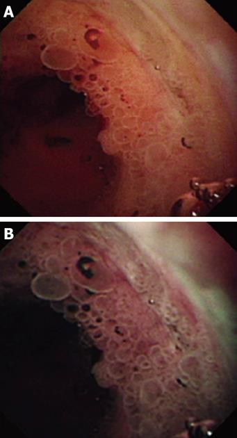
Figure 1 Peroral pancreatoscopy images.
A: Peroral pancreatoscopy of the main pancreatic duct demonstrating the presence of papillary tumor; small, ovoid papillary projections can be seen; B: The same projections are pictures here under observation with narrow band imaging; the surface structure of the lesions is much better visualized. (The figure is from Itoi et al[20] and reproduced with permission from Elsevier Inc.)
- Citation: Turner BG, Brugge WR. Diagnostic and therapeutic endoscopic approaches to intraductal papillary mucinous neoplasm. World J Gastrointest Surg 2010; 2(10): 337-341
- URL: https://www.wjgnet.com/1948-9366/full/v2/i10/337.htm
- DOI: https://dx.doi.org/10.4240/wjgs.v2.i10.337


