Copyright
©The Author(s) 2023.
World J Gastrointest Surg. Mar 27, 2023; 15(3): 488-494
Published online Mar 27, 2023. doi: 10.4240/wjgs.v15.i3.488
Published online Mar 27, 2023. doi: 10.4240/wjgs.v15.i3.488
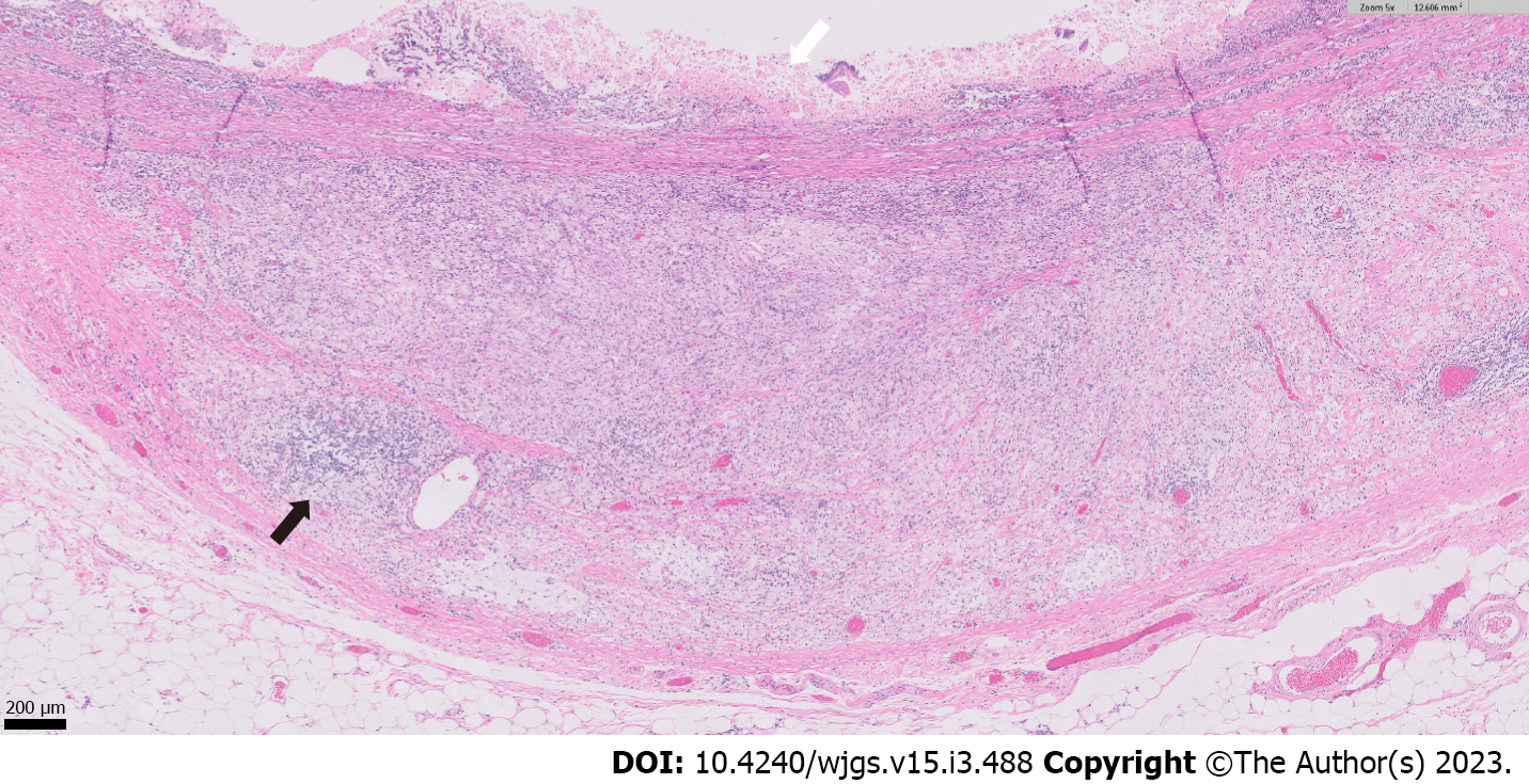
Figure 5 High-magnification section of the specimen.
Higher magnification (5 × objective magnification) image showing mucosal ulceration (white arrow) with abundant infiltration of the underlying small intestinal wall by collections of foamy, lipid-laden histiocytes (xanthogranulomatous inflammation), along with lymphocytes and plasma cells (example highlighted by black arrow).
- Citation: Wang W, Korah M, Bessoff KE, Shen J, Forrester JD. Xanthogranulomatous inflammation requiring small bowel anastomosis revision: A case report. World J Gastrointest Surg 2023; 15(3): 488-494
- URL: https://www.wjgnet.com/1948-9366/full/v15/i3/488.htm
- DOI: https://dx.doi.org/10.4240/wjgs.v15.i3.488


