Copyright
©The Author(s) 2023.
World J Gastrointest Surg. Mar 27, 2023; 15(3): 488-494
Published online Mar 27, 2023. doi: 10.4240/wjgs.v15.i3.488
Published online Mar 27, 2023. doi: 10.4240/wjgs.v15.i3.488
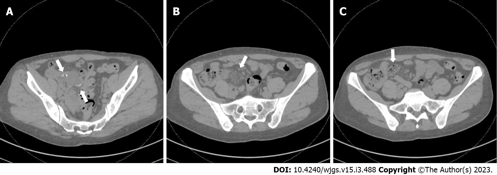
Figure 1 Computed tomography scan of her abdomen and pelvis without contrast.
A: Prior anastomotic suture line is visualized (arrows) and appeared intact; B: Mild surrounding inflammatory stranding (arrow) seen near the prior anastomosis; C: Fecalization (arrow) noted in small bowel adjacent to anastomosis.
- Citation: Wang W, Korah M, Bessoff KE, Shen J, Forrester JD. Xanthogranulomatous inflammation requiring small bowel anastomosis revision: A case report. World J Gastrointest Surg 2023; 15(3): 488-494
- URL: https://www.wjgnet.com/1948-9366/full/v15/i3/488.htm
- DOI: https://dx.doi.org/10.4240/wjgs.v15.i3.488


