Copyright
©The Author(s) 2015.
World J Diabetes. Feb 15, 2015; 6(1): 175-183
Published online Feb 15, 2015. doi: 10.4239/wjd.v6.i1.175
Published online Feb 15, 2015. doi: 10.4239/wjd.v6.i1.175
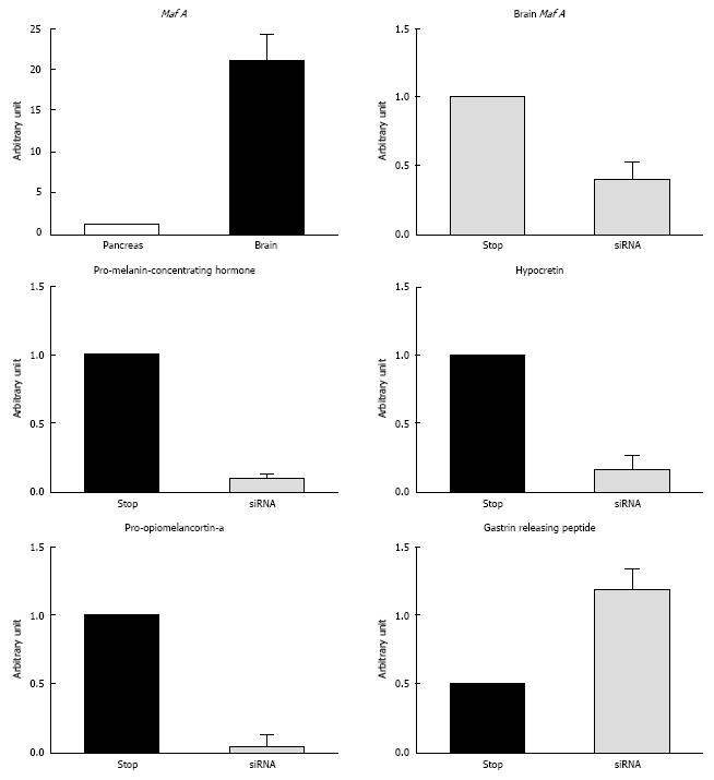
Figure 3 Suppression of MafA mRNA by siRNA in the brain and resulting alternation of related genes.
A designed small interfering RNA (siRNA) oligomer for mouse Mafa was intravenously injected using the hydrodynamic method according to a procedure described by Hamar et al[58]. A DNA microarray analysis was then performed using Affymetrix GeneChip technology. The mRNA levels were quantified using real-time PCR. The details of the experiment have been described previously. Expression level of Mafa mRNA in the brain. The expression level of Mafa mRNA in the brain was 20 times higher than that of Mafa mRNA in the pancreas, as assessed using real-time PCR. Suppression of Mafa in mice using siRNA in the brain. The mRNA expression level take out in the brain tissue are shown. The Mafa mRNA expression level was significantly downregulated by the siRNA. Pro-melanin-concentrating hormone, Hypocretin, and Pro-opiomelanocortin-a were downregulated, and Gastrin-releasing peptide was upregulated, as assessed using real-time PCR with specific primers.
- Citation: Tsuchiya M, Misaka R, Nitta K, Tsuchiya K. Transcriptional factors, Mafs and their biological roles. World J Diabetes 2015; 6(1): 175-183
- URL: https://www.wjgnet.com/1948-9358/full/v6/i1/175.htm
- DOI: https://dx.doi.org/10.4239/wjd.v6.i1.175


