Copyright
©The Author(s) 2017.
World J Gastrointest Oncol. Feb 15, 2017; 9(2): 50-61
Published online Feb 15, 2017. doi: 10.4251/wjgo.v9.i2.50
Published online Feb 15, 2017. doi: 10.4251/wjgo.v9.i2.50
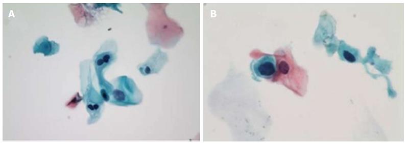
Figure 1 Cytology of anal intraepithelial lesions.
A: LSIL, with representative binucleate hyperchromatic cells (koilocytes) and nuclear enlargement (Papanicolaou stain, original magnification × 400); B: HSIL, with representative markedly increased nuclear to cytoplasmis ratio as comparted to LSIL at left (Papanicolaou stain, oil immersion, original magnification × 1000). Reproduced with permission[85]. LSIL: Low grade squamous intraepithelial lesions; HSIL: High grade squamous intraepithelial lesions.
- Citation: Roberts JR, Siekas LL, Kaz AM. Anal intraepithelial neoplasia: A review of diagnosis and management. World J Gastrointest Oncol 2017; 9(2): 50-61
- URL: https://www.wjgnet.com/1948-5204/full/v9/i2/50.htm
- DOI: https://dx.doi.org/10.4251/wjgo.v9.i2.50


