Copyright
©The Author(s) 2023.
World J Gastrointest Oncol. Jul 15, 2023; 15(7): 1119-1134
Published online Jul 15, 2023. doi: 10.4251/wjgo.v15.i7.1119
Published online Jul 15, 2023. doi: 10.4251/wjgo.v15.i7.1119
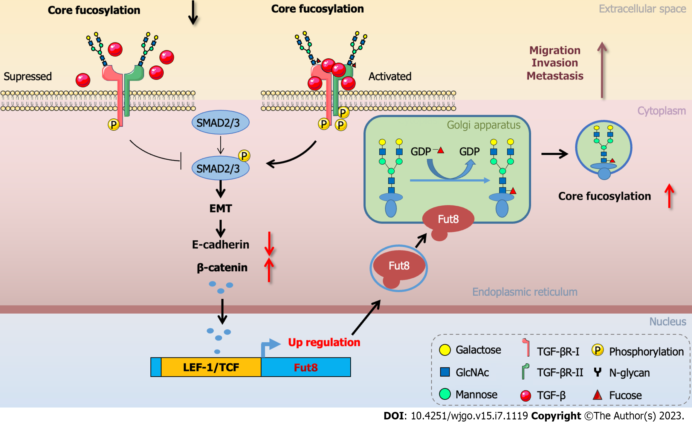
Figure 3 Core fucosylation modulated epithelial to mesenchymal transition during cancer progression.
Loss of core fucosylation impaired the binding of transforming growth factor β to its receptor and inhibited the phosphorylation of regulatory Smad2 and Smad3. During epithelial to mesenchymal transition, the expression of E-cadherin was suppressed, resulting in the accumulation of β-catenin in the nucleus, where it complexed with lymphoid enhancer-binding factor 1 to transactivate fucosyltransferase 8 expression. The core fucosylation level on surface molecules of cancer cells was increased which promoted cancer cell malignant biological properties involving migration, invasion, and metastasis. EMT: Epithelial to mesenchymal transition; TCF: T cell factor; LEF: Lymphoid enhancer-binding factor 1; TGF: Transforming growth factor. This figure was summarized and modified from Tu et al[46], Chen et al[74], and Lin et al[101].
- Citation: Zhang NZ, Zhao LF, Zhang Q, Fang H, Song WL, Li WZ, Ge YS, Gao P. Core fucosylation and its roles in gastrointestinal glycoimmunology. World J Gastrointest Oncol 2023; 15(7): 1119-1134
- URL: https://www.wjgnet.com/1948-5204/full/v15/i7/1119.htm
- DOI: https://dx.doi.org/10.4251/wjgo.v15.i7.1119


