Copyright
©The Author(s) 2021.
World J Gastrointest Oncol. Jun 15, 2021; 13(6): 509-535
Published online Jun 15, 2021. doi: 10.4251/wjgo.v13.i6.509
Published online Jun 15, 2021. doi: 10.4251/wjgo.v13.i6.509
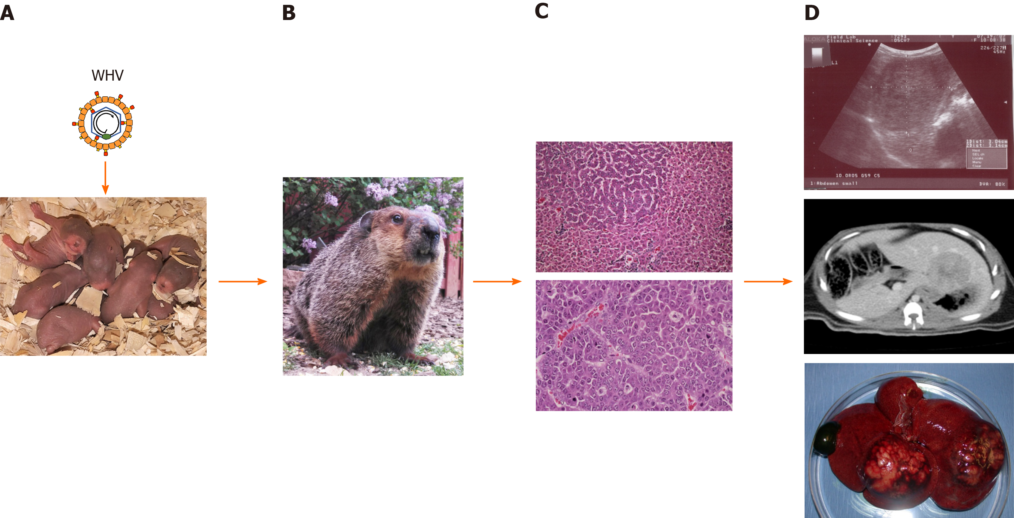
Figure 1 Schematic presentation of woodchuck hepatitis virus-induced liver disease progression and detection of tumors within woodchuck liver.
A: Neonatal woodchucks are experimentally infected with woodchuck hepatitis virus (WHV) to model vertical hepatitis B virus transmission in humans; B: WHV infection progresses to chronic hepatitis B in adult woodchucks after approximately 1 year; C: Chronic WHV carrier woodchucks develop liver tumors during the next 1-1½ years. A focus of altered hepatocytes (FAH) in liver (top) and an undifferentiated liver tumor (bottom) are shown; D: Localization of liver tumors by ultrasonography (top) and computed tomography (middle). The liver of a woodchuck with two larger hepatocellular neoplasms (HCC) is shown (bottom). With permission from Elsevier, pictures shown in C were reprinted from: Tennant BC, Toshkov IA, Peek SF, Jacob JR, Menne S, Hornbuckle WE, Schinazi RD, Korba BE, Cote PJ, Gerin JL. Hepatocellular carcinoma in the woodchuck model of hepatitis B virus infection. Gastroenterology 2004; 127(5): S283-S293. Copyright ©American Gastroenterological Association 2004. Published by Elsevier[37]. WHV: Woodchuck hepatitis virus.
- Citation: Suresh M, Menne S. Application of the woodchuck animal model for the treatment of hepatitis B virus-induced liver cancer. World J Gastrointest Oncol 2021; 13(6): 509-535
- URL: https://www.wjgnet.com/1948-5204/full/v13/i6/509.htm
- DOI: https://dx.doi.org/10.4251/wjgo.v13.i6.509


