Copyright
©The Author(s) 2017.
World J Gastrointest Endosc. Mar 16, 2017; 9(3): 139-144
Published online Mar 16, 2017. doi: 10.4253/wjge.v9.i3.139
Published online Mar 16, 2017. doi: 10.4253/wjge.v9.i3.139
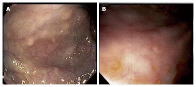
Figure 2 Gross appearance of colonic lesions on endoscopy.
Wide (A) and close-up (B) views of approximately 5 mm whitish-yellowish polypoid lesions found throughout the colon. Biopsy and immunohistochemical staining demonstrated CD1a, S-100, and langerin reactivity confirming langerhans cell histiocytosis-related lesions (see Figure 3).
- Citation: Karimzada MM, Matthews MN, French SW, DeUgarte D, Kim DY. Langerhans cell histiocytosis masquerading as acute appendicitis: Case report and review. World J Gastrointest Endosc 2017; 9(3): 139-144
- URL: https://www.wjgnet.com/1948-5190/full/v9/i3/139.htm
- DOI: https://dx.doi.org/10.4253/wjge.v9.i3.139


