Copyright
©The Author(s) 2016.
World J Gastrointest Endosc. Nov 16, 2016; 8(19): 690-696
Published online Nov 16, 2016. doi: 10.4253/wjge.v8.i19.690
Published online Nov 16, 2016. doi: 10.4253/wjge.v8.i19.690
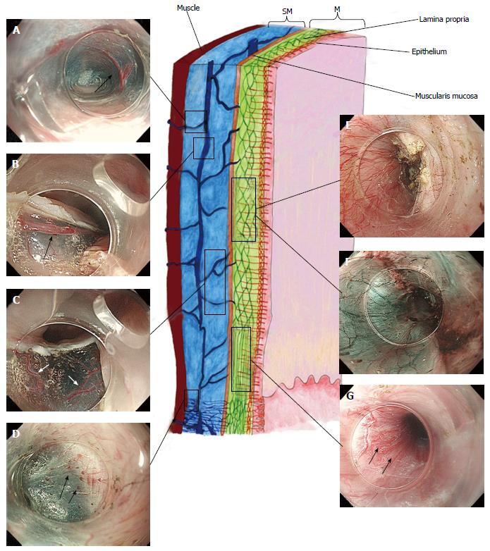
Figure 2 Esophageal wall and esophago-gastric junction vasculature: Schematic illustration and endoscopic corresponding images (high magnification images).
Black arrow indicates vessels. This image was originally published in “Treatment Strategies Gastroenterology”[28]. A: Perforating vessels from the outer esophagus to the submucosal vessel; image captured during tunnelization in POEM (bottom side muscle layer, left side submucosal lifting); B: Submucosal drainage vessel (mucosal layer lifted on during ESD). These veins can become esophageal varices in portal hypertension; C: Submucosal vessels connecting the drainage veins to the mucosal branching vessels (in the lamina propria); D: Spindle veins immediately below the GEJ (in left side of the image, in blue, the submucosa and in the right side the muscle); E and F: Whitet light and NBI of the branching vessels (seen from inside the submucosal tunnel). Backside of the mucosa on the left; muscle-already cut-on the right; G: Passage between lower esophagus and GEJ. In the image is possible to recognize, in different planes, all the vessel of the submucosa and lamina propria (palisade vessels). POEM: Per-oral endoscopic myotomy; ESD: Endoscopic submucosal dissection; GEJ: Esophagogastric junction; NBI: Narrow band imaging; M: Mucosa; SM: Submucosa.
- Citation: Maselli R, Inoue H, Ikeda H, Onimaru M, Yoshida A, Santi EG, Sato H, Hayee B, Kudo SE. Microvasculature of the esophagus and gastroesophageal junction: Lesson learned from submucosal endoscopy. World J Gastrointest Endosc 2016; 8(19): 690-696
- URL: https://www.wjgnet.com/1948-5190/full/v8/i19/690.htm
- DOI: https://dx.doi.org/10.4253/wjge.v8.i19.690


