Copyright
©The Author(s) 2015.
World J Gastrointest Endosc. Mar 16, 2015; 7(3): 192-205
Published online Mar 16, 2015. doi: 10.4253/wjge.v7.i3.192
Published online Mar 16, 2015. doi: 10.4253/wjge.v7.i3.192
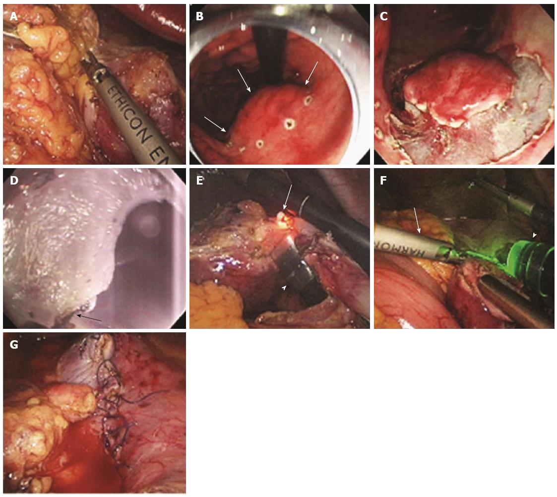
Figure 7 Procedure for laparoscopy-assisted endoscopic full-thickness resection.
A: Laparoscopic view while the lesser omentum attached around the tumor site was dissected; B: Endoscopic view after marking around the gastric subepithelial tumor (white arrows) located on the lesser curvature side of the gastric body; C: Gastroscopic view after incision as deep as the submucosal layer around the lesion; D: Gastroscopic view of the full-thickness incision from inside the stomach using the IT knife (white arrow); E: Laparoscopic view of the full-thickness incision from inside the stomach using the IT knife (arrow, the tip of the IT knife; arrowhead, the gastroscope); F: Laparoscopic view of the remaining full-thickness incision from outside the stomach using a Harmonic ACE (arrow); G: Laparoscopic view after laparoscopic hand-sewn closure of the gastric-wall defect (adopted from Abe et al[58]).
- Citation: Kim HH. Endoscopic treatment for gastrointestinal stromal tumor: Advantages and hurdles. World J Gastrointest Endosc 2015; 7(3): 192-205
- URL: https://www.wjgnet.com/1948-5190/full/v7/i3/192.htm
- DOI: https://dx.doi.org/10.4253/wjge.v7.i3.192


