Copyright
©2013 Baishideng Publishing Group Co.
World J Gastrointest Endosc. Nov 16, 2013; 5(11): 540-550
Published online Nov 16, 2013. doi: 10.4253/wjge.v5.i11.540
Published online Nov 16, 2013. doi: 10.4253/wjge.v5.i11.540
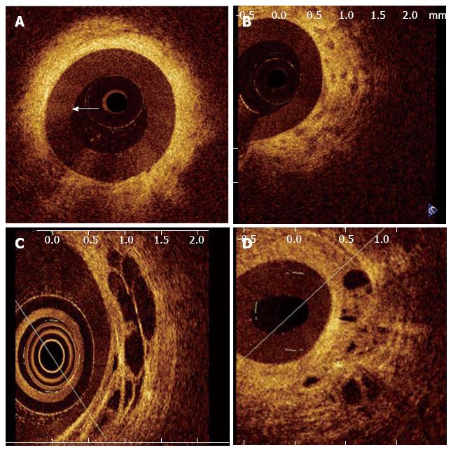
Figure 7 Optical coherence tomography image of a patient with a benign stricture.
The three-layered structure of the biliary wall is recognizable (Color online). A-D shows images of malignant bile duct strictures. B: Disorganized layered structure with unidentifiable margins and a strongly heterogeneous back-scattering signal; C: Large, nonreflective areas in the intermediate layer suggesting the tumor vessels; D: Malignant stricture due to hilar metastases of an esophageal squamous carcinoma showing nonreflective areas and disorganized layer architecture[2].
- Citation: Mahmud MS, May GR, Kamal MM, Khwaja AS, Sun C, Vitkin A, Yang VX. Imaging pancreatobiliary ductal system with optical coherence tomography: A review. World J Gastrointest Endosc 2013; 5(11): 540-550
- URL: https://www.wjgnet.com/1948-5190/full/v5/i11/540.htm
- DOI: https://dx.doi.org/10.4253/wjge.v5.i11.540


