Copyright
©2013 Baishideng Publishing Group Co.
World J Gastrointest Endosc. Nov 16, 2013; 5(11): 540-550
Published online Nov 16, 2013. doi: 10.4253/wjge.v5.i11.540
Published online Nov 16, 2013. doi: 10.4253/wjge.v5.i11.540
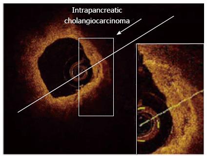
Figure 6 Adenocarcinoma (neoplasia) of the common bile duct at early stage, detected with optical coherence tomography probe maintained inside the endoscopic retrograde cholangiopancheatography catheter.
In the presence of Adenocarcinoma (neoplasia), optical coherence tomography (OCT) patterns showed distorted common bile duct (CBD) wall structure (Color online). All three-layer architecture and their linear and regular surface, normally giving a homogeneous back-scattered signal, are not recognizable. OCT image shows heterogeneous back-scattered signal with minute, multiple, nonreflective areas (necrotic areas) in the highly disorganized CBD microstructure. Therefore, epithelial structure and various biliary disorders in early-stage of cancer can be distinguishable with OCT[67].
- Citation: Mahmud MS, May GR, Kamal MM, Khwaja AS, Sun C, Vitkin A, Yang VX. Imaging pancreatobiliary ductal system with optical coherence tomography: A review. World J Gastrointest Endosc 2013; 5(11): 540-550
- URL: https://www.wjgnet.com/1948-5190/full/v5/i11/540.htm
- DOI: https://dx.doi.org/10.4253/wjge.v5.i11.540


