Copyright
©2012 Baishideng Publishing Group Co.
World J Gastrointest Endosc. Apr 16, 2012; 4(4): 151-156
Published online Apr 16, 2012. doi: 10.4253/wjge.v4.i4.151
Published online Apr 16, 2012. doi: 10.4253/wjge.v4.i4.151
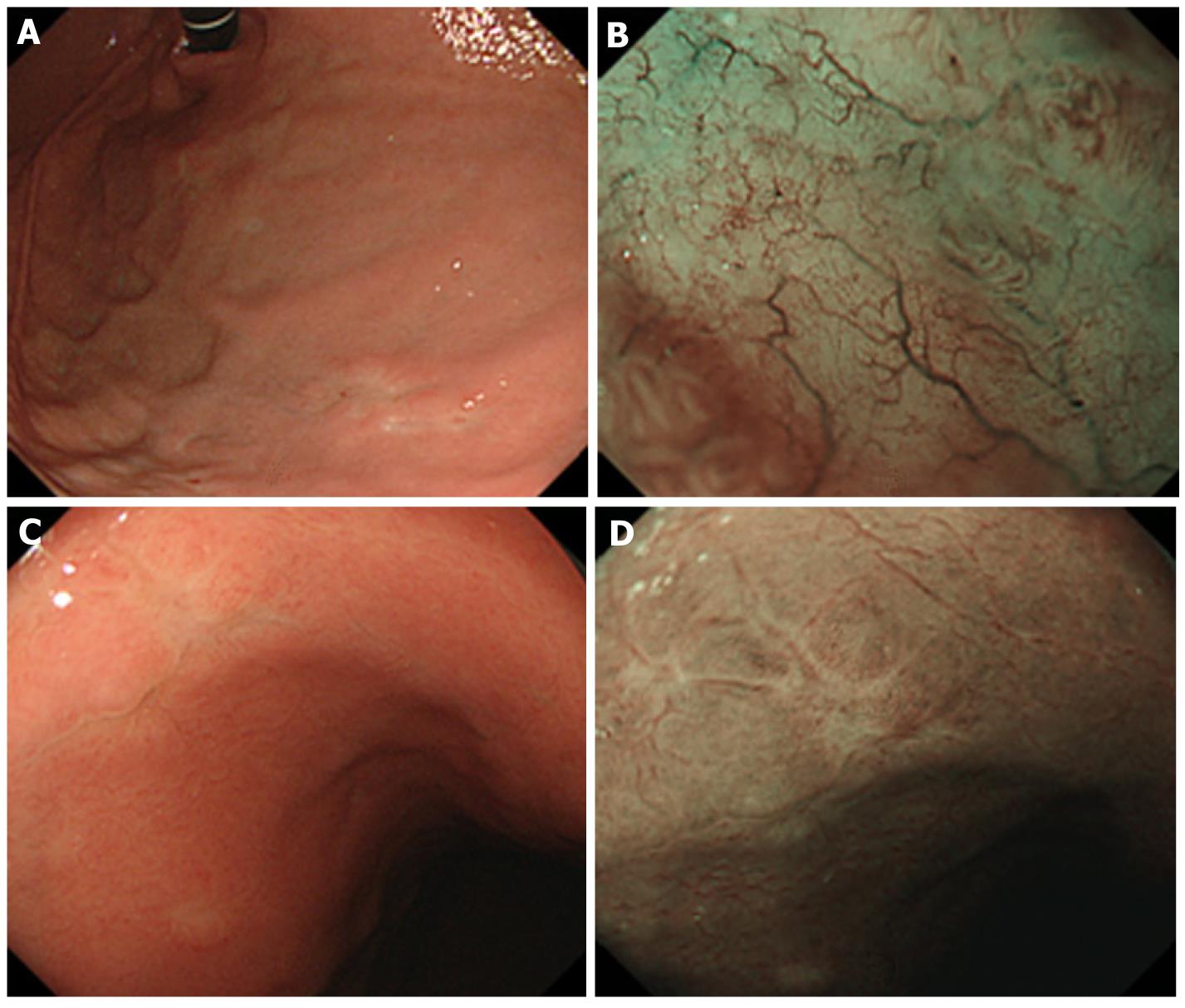
Figure 1 Routine observation revealed multiple ill-delineated brownish depressions in the stomach.
A: A brownish depression in the anterior wall of the upper gastric corpus; B: Narrow band imaging (NBI) findings of Figure 1A (TLA+); C: A brownish depression in the anterior wall of the middle portion of the gastric corpus; D: NBI findings of Figure 1C (TLA-). TLA: Tree-like appearance.
- Citation: Nonaka K, Ishikawa K, Arai S, Nakao M, Shimizu M, Sakurai T, Nagata K, Nishimura M, Togawa O, Ochiai Y, Sasaki Y, Kita H. A case of gastric mucosa-associated lymphoid tissue lymphoma in which magnified endoscopy with narrow band imaging was useful in the diagnosis. World J Gastrointest Endosc 2012; 4(4): 151-156
- URL: https://www.wjgnet.com/1948-5190/full/v4/i4/151.htm
- DOI: https://dx.doi.org/10.4253/wjge.v4.i4.151


