Copyright
©2012 Baishideng Publishing Group Co.
World J Gastrointest Endosc. Mar 16, 2012; 4(3): 57-64
Published online Mar 16, 2012. doi: 10.4253/wjge.v4.i3.57
Published online Mar 16, 2012. doi: 10.4253/wjge.v4.i3.57
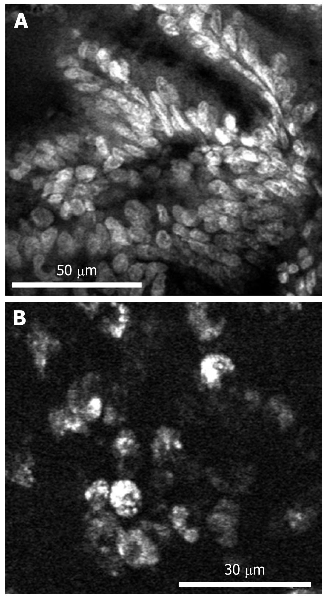
Figure 3 Imaging of vascular endothelial growth factor in the biopsy specimen of human colorectal adenocarcinoma.
A: Nonspecific nuclear and cellular staining using acriflavine; B: VEGF-specific staining using AF488-labeled antibodies. The antibody accumulates in the cytoplasm of the tumor cells, but not the nuclei. Reproduced with permission from Foersch et al[39].
- Citation: Kwon YS, Cho YS, Yoon TJ, Kim HS, Choi MG. Recent advances in targeted endoscopic imaging: Early detection of gastrointestinal neoplasms. World J Gastrointest Endosc 2012; 4(3): 57-64
- URL: https://www.wjgnet.com/1948-5190/full/v4/i3/57.htm
- DOI: https://dx.doi.org/10.4253/wjge.v4.i3.57


