Copyright
©The Author(s) 2019.
World J Gastrointest Endosc. Jan 16, 2019; 11(1): 5-21
Published online Jan 16, 2019. doi: 10.4253/wjge.v11.i1.5
Published online Jan 16, 2019. doi: 10.4253/wjge.v11.i1.5
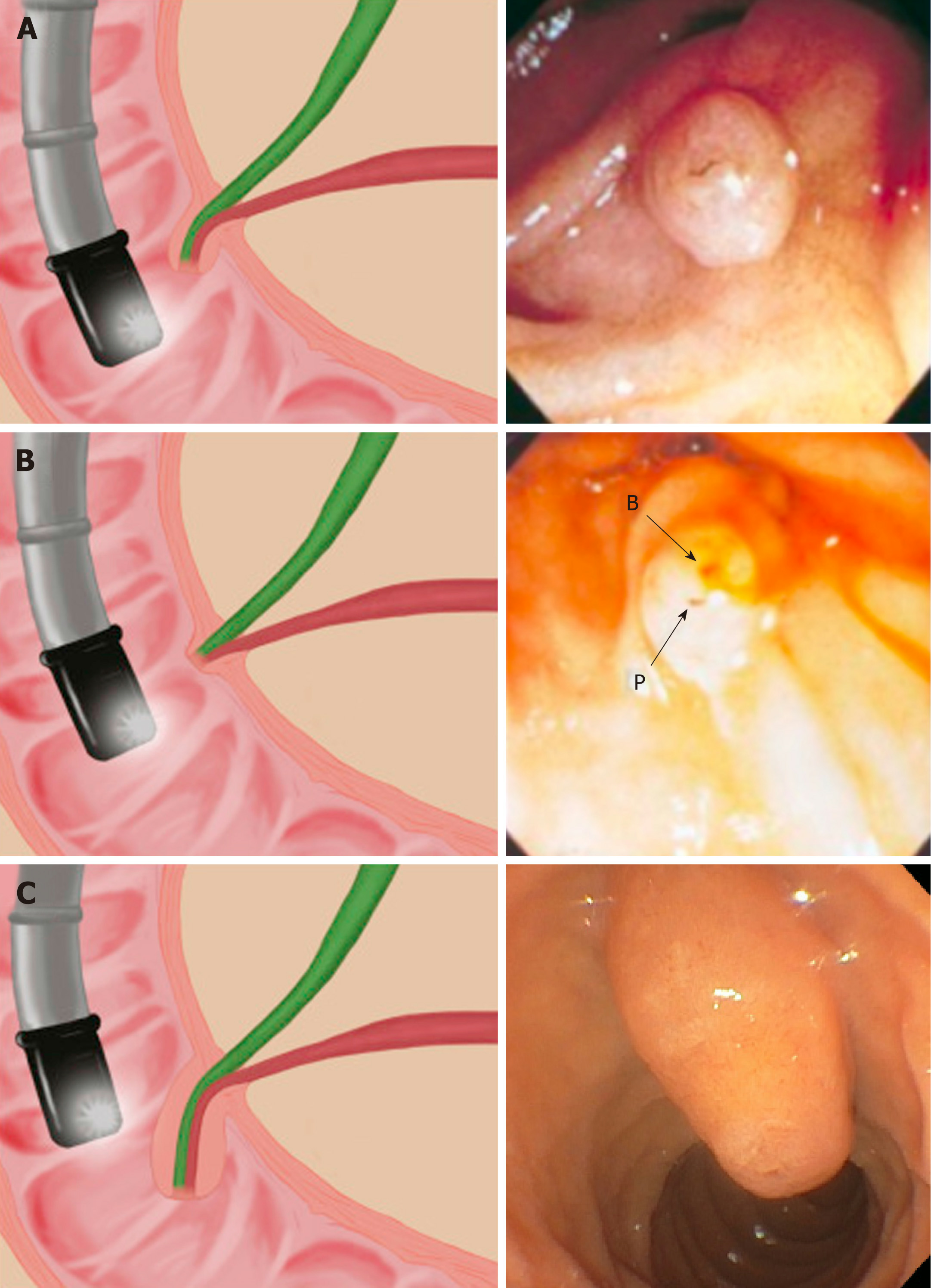
Figure 3 Drawing and corresponding endoscopic view of anatomic variants seen during endoscopic retrograde cholangiopancreatography of the ampulla and major duodenal papilla.
A: Normal ampulla and pancreatobiliary junction; B: No common channel; endoscopically, two separate openings (P: Pancreatic duct; and B: Bile duct) may be seen at the papillary tip; C: Large, protuberant, and/or redundant papilla[6,7].
- Citation: Berry R, Han JY, Tabibian JH. Difficult biliary cannulation: Historical perspective, practical updates, and guide for the endoscopist. World J Gastrointest Endosc 2019; 11(1): 5-21
- URL: https://www.wjgnet.com/1948-5190/full/v11/i1/5.htm
- DOI: https://dx.doi.org/10.4253/wjge.v11.i1.5


