Copyright
©The Author(s) 2016.
World J Hepatol. Mar 18, 2016; 8(8): 411-420
Published online Mar 18, 2016. doi: 10.4254/wjh.v8.i8.411
Published online Mar 18, 2016. doi: 10.4254/wjh.v8.i8.411
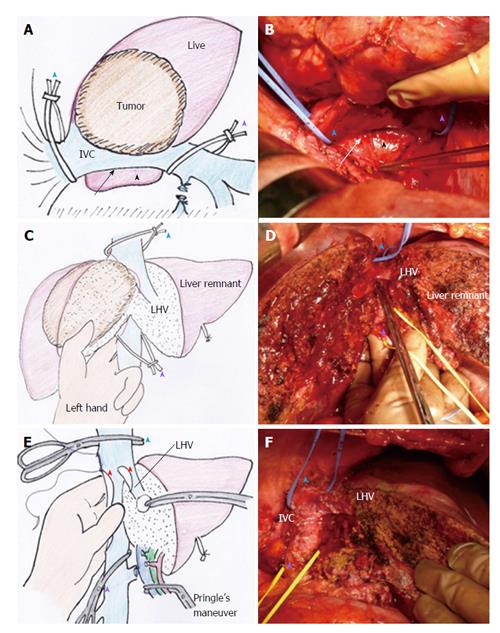
Figure 2 Retrocaval liver lifting maneuver, performed in case 2.
A (illustration) and B (intraoperative image): The retrocaval space (arrow) was dissected broadly from the right lateral view toward the inner aspect of Spiegel’s lobe (black arrowhead), after which the supra- and infrahepatic IVC were taped to prepare cross-clamping for THVE (blue arrowhead and purple arrowhead); C (illustration) and D (intraoperative image): The index and middle fingers of the surgeon’s left hand were placed into the dissected retrocaval space, and the IVC was compressed ventrally to control bleeding around the IVC during deep parenchymal transection, after which the thumb finger of the surgeon’s left hand was used to spread the transection plane of the liver (C, which also shows the tumor status); D: Hepatic parenchymal transection is completed, except for the IVC involved site, before applying THVE; E (illustration) and F (intraoperative image): The liver specimen was excised along with the involved IVC and LHV wall (red arrowheads), and the backflow bleeding from the cut orifice of IVC was controlled by pinching the IVC using the surgeon’s left hand from its placement in the retrocaval space; F shows the view after reconstruction of IVC and LHV. See Figure 1B for tumor status. IVC: Inferior vena cava; LHV: Left hepatic vein; THVE: Total hepatic vascular exclusion.
- Citation: Ko S, Kirihataya Y, Matsumoto Y, Takagi T, Matsusaka M, Mukogawa T, Ishikawa H, Watanabe A. Retrocaval liver lifting maneuver and modifications of total hepatic vascular exclusion for liver tumor resection. World J Hepatol 2016; 8(8): 411-420
- URL: https://www.wjgnet.com/1948-5182/full/v8/i8/411.htm
- DOI: https://dx.doi.org/10.4254/wjh.v8.i8.411


