Copyright
©The Author(s) 2016.
World J Hepatol. Nov 28, 2016; 8(33): 1419-1441
Published online Nov 28, 2016. doi: 10.4254/wjh.v8.i33.1419
Published online Nov 28, 2016. doi: 10.4254/wjh.v8.i33.1419
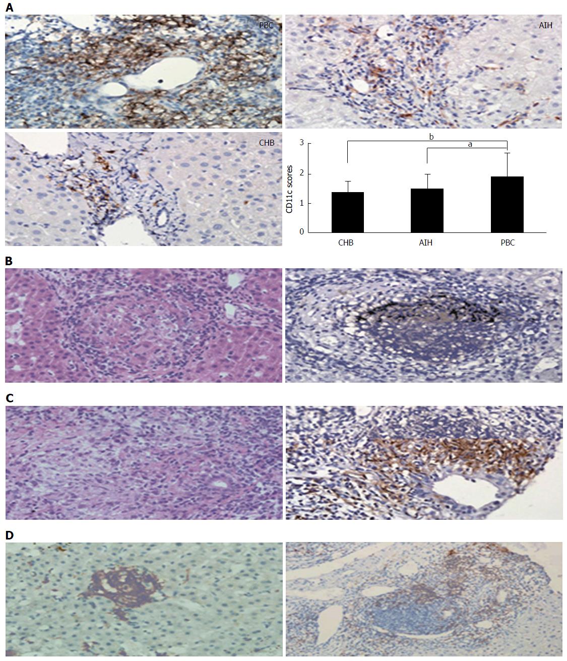
Figure 2 Histology (hematoxylin and eosin staining) and immunochemical staining of primary biliary cholangitis.
A: Magnification × 400; B: Magnification × 400; C, D: Magnification × 400 (left); Magnification × 200 (right). PBC livers demonstrated observably stronger portal area immunostain for CD11c, the position of CD11c sedimentation scored on a 0-4 scale to be compared among PBC, AIH and CHB patients (aP < 0.05, bP < 0.01) (A); PBC hepatic granulomatous lesions were classically situated within portal areas, generally near or around the impaired bile duct (B); Hepatic granulomatous lesions were also occasionally detected in the liver lobule or close to the germinal center (C)[137]. PBC: Primary biliary cholangitis; AIH: Autoimmune hepatitides; CHB: Chronic hepatitis B.
- Citation: Huang YQ. Recent advances in the diagnosis and treatment of primary biliary cholangitis. World J Hepatol 2016; 8(33): 1419-1441
- URL: https://www.wjgnet.com/1948-5182/full/v8/i33/1419.htm
- DOI: https://dx.doi.org/10.4254/wjh.v8.i33.1419


