Copyright
©The Author(s) 2016.
World J Hepatol. Jul 8, 2016; 8(19): 796-814
Published online Jul 8, 2016. doi: 10.4254/wjh.v8.i19.796
Published online Jul 8, 2016. doi: 10.4254/wjh.v8.i19.796
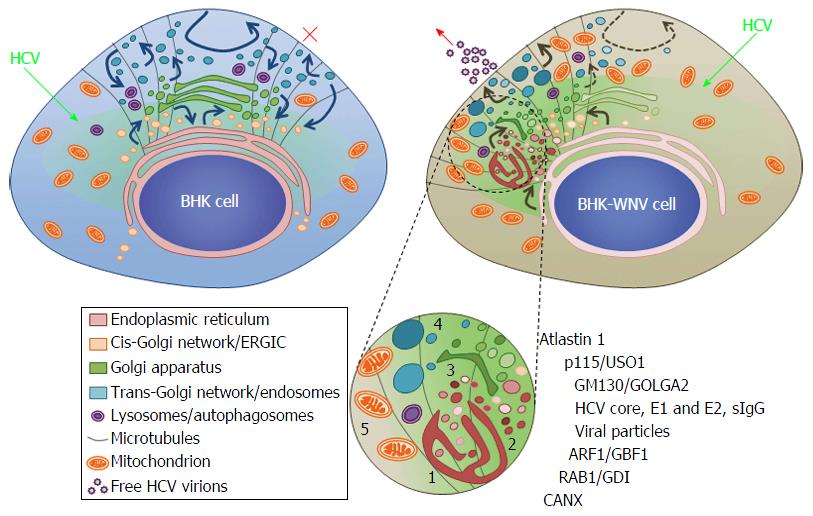
Figure 14 Model of a hepatitis C virus assembly compartment in baby hamster kidney-West Nile virus cells.
Schematic organization of cell traffic in parental BHK-21 (top left) and BHK-WNV (top right) cells; curved arrows represent cell traffic; HCV genome is produced by the P2B plasmid system (green arrows) and expressed (greenish areas) in the cytoplasm of BHK cells. Bottom, sketches’ legend and close up of a HCV assembly site (cf. also Figure 2B): (1) convoluted membranes; (2) vesicular packets; (3) Golgi cisternæ; (4) large vesicles filled with viral particles; and (5) mitochondrion; host and viral factors identified within this compartment. SIgG: Antibodies from the serum of a cured HCV patient; CANX: Calnexin; HCV: Hepatitis C virus; BHK-WNV: Baby hamster kidney-West Nile virus; ERGIC: Endoplasmic reticulum-Golgi intermediate compartment; GDI: GDP dissociation inhibitor.
- Citation: Triyatni M, Berger EA, Saunier B. Assembly and release of infectious hepatitis C virus involving unusual organization of the secretory pathway. World J Hepatol 2016; 8(19): 796-814
- URL: https://www.wjgnet.com/1948-5182/full/v8/i19/796.htm
- DOI: https://dx.doi.org/10.4254/wjh.v8.i19.796


