Copyright
©The Author(s) 2016.
World J Hepatol. Jul 8, 2016; 8(19): 796-814
Published online Jul 8, 2016. doi: 10.4254/wjh.v8.i19.796
Published online Jul 8, 2016. doi: 10.4254/wjh.v8.i19.796
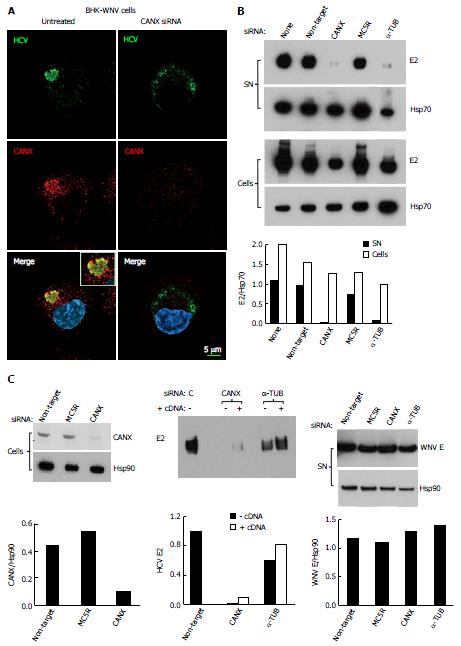
Figure 3 Involvement of calnexin and alpha-tubulin in the release of hepatitis C virus particles by baby hamster kidney-West Nile virus cells.
A: BHK-WNV cells were treated, or not, with CANX siRNA and, three days later, transfected with the HCV-coding and P2B plasmids. Two days later, IF was performed with anti-HCV serum of Figure 1 (green) and anti-CANX antibody (red) followed by confocal microcopy analysis; nucleus were counterstained with DAPI (blue); B: Top panels: BHK-WNV cells were treated with the indicated siRNA for 2 d, then transfected with a plasmid encoding full length HCV (HCVbp) in the cytoplasm. Contents in E2 envelope protein of both supernatant (SN) and cell lysate (Cells) were analyzed 2 d later by Western blot (WB); Bottom panel: Densitometry analysis; C: Left panels: BHK-WNV cells were treated with the indicated siRNA for 2 d and content in CANX was analyzed by WB; Hsp90 was used as a control; Middle panel: BHK-WNV cells were treated with siRNA targeting CANX or a-TUB transcript for 2 d; cells were then reseeded and transfected the next day with the HCV-coding plasmid together with a control plasmid (-) or one expressing the cDNA of the knocked-down transcript (+). Two days later, HCV materials released in SN were analyzed by WB; Right panels: BHK-WNV treated as in (B) were transfected with a plasmid encoding West Nile virus (WNV) structural genes (core, prM and E). Two days later, materials released in the SN were analyzed by WB with an antibody recognizing WNV E (29); Hsp90 was used as a control; Bottom panels: Densitometry analyses. CANX: Calnexin; α-TUB: Alpha-tubulin; HCV: Hepatitis C virus; BHK-WNV: Baby hamster kidney-West Nile virus; C: Control siRNA.
- Citation: Triyatni M, Berger EA, Saunier B. Assembly and release of infectious hepatitis C virus involving unusual organization of the secretory pathway. World J Hepatol 2016; 8(19): 796-814
- URL: https://www.wjgnet.com/1948-5182/full/v8/i19/796.htm
- DOI: https://dx.doi.org/10.4254/wjh.v8.i19.796


