Copyright
©The Author(s) 2015.
World J Hepatol. Oct 18, 2015; 7(23): 2432-2448
Published online Oct 18, 2015. doi: 10.4254/wjh.v7.i23.2432
Published online Oct 18, 2015. doi: 10.4254/wjh.v7.i23.2432
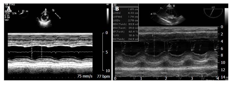
Figure 7 Transesophageal measurements of fractional shortening.
A: TG Mid SAX view of the left ventricle showing M-mode measurement of LVEDD and LVESD normalized for LVEDD. The bidimensional image is usually best imaged at a multiplane angle of 0°; B: TG LAX view of left ventricle showing M-Mode measurement of LVEDD and LVESD normalized for LVEDD usually best imaged at an angle of approximately 80°-110°. LAX: Long axis view; TG: Transgastric; LVEDD: Left ventricle end diastolic diameter; LVESD: Left ventricle end systolic diameter. Modified by Guarracino Fabio et al[61].
- Citation: De Pietri L, Mocchegiani F, Leuzzi C, Montalti R, Vivarelli M, Agnoletti V. Transoesophageal echocardiography during liver transplantation. World J Hepatol 2015; 7(23): 2432-2448
- URL: https://www.wjgnet.com/1948-5182/full/v7/i23/2432.htm
- DOI: https://dx.doi.org/10.4254/wjh.v7.i23.2432


