Copyright
©2014 Baishideng Publishing Group Inc.
World J Hepatol. Aug 27, 2014; 6(8): 580-595
Published online Aug 27, 2014. doi: 10.4254/wjh.v6.i8.580
Published online Aug 27, 2014. doi: 10.4254/wjh.v6.i8.580
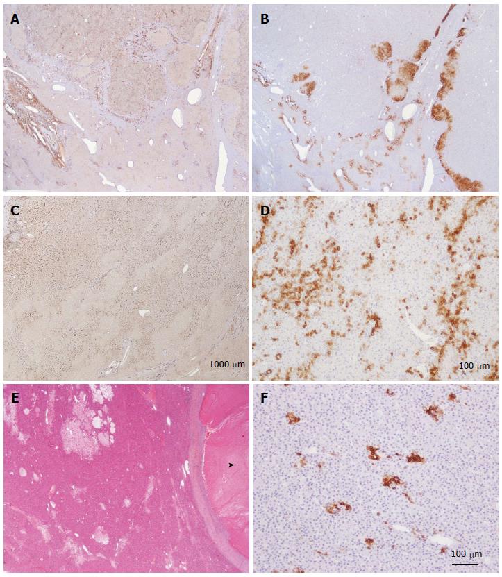
Figure 19 Unclassified hepatocellular adenoma.
A, B: Woman born in 1988; oral contraceptives for 4 years. BMI 16.8. Abdominal pain. Imaging: one nodule 11 cm. No final diagnosis. Left hepatectomy 2009. A: Diffuse expression of CD34 in hepatocellular adenoma (HCA) (top) contrasting with adjacent non tumoral liver (below). B: No expression of glutamine synthase (GS) except at the periphery of the HCA. Here the nodule is divided in 2 parts at its periphery by a thin band of normal tissue containing vessels. C, D: Woman born in 1975; oral contraceptives for 12 years. BMI 24.2 kg/m2. Hemorrhage. Imaging: one nodule 5 cm, HCA. Right hepatectomy 2003. C: Widespread but not diffuse expression of CD34 within the HCA. D: Numerous cells overexpressing CK7: small cells looking like progenitor cells and intermediate cells. GS was normal. E, F: Same patient as Figure 5I. E: HE: thick fibrous rim around a necrotic area (arrowhead); peliotic areas within the viable HCA. F: Some small CK7 positive cells dispersed within the HCA. GS (not shown) was normal. CD34 staining was more or less diffuse (not shown).
- Citation: Sempoux C, Balabaud C, Bioulac-Sage P. Pictures of focal nodular hyperplasia and hepatocellular adenomas. World J Hepatol 2014; 6(8): 580-595
- URL: https://www.wjgnet.com/1948-5182/full/v6/i8/580.htm
- DOI: https://dx.doi.org/10.4254/wjh.v6.i8.580


