Copyright
©2014 Baishideng Publishing Group Inc.
World J Hepatol. Aug 27, 2014; 6(8): 580-595
Published online Aug 27, 2014. doi: 10.4254/wjh.v6.i8.580
Published online Aug 27, 2014. doi: 10.4254/wjh.v6.i8.580
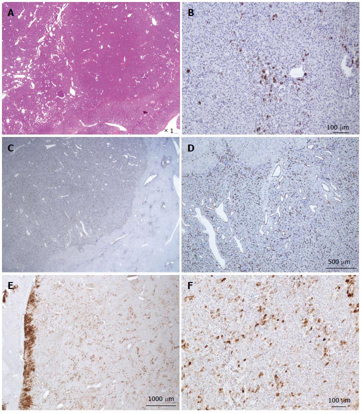
Figure 18 β-catenin hepatocellular adenoma.
A, F: Same patient as Figure 5G. A: HE: Numerous vessels dispersed within the hepatocellular proliferation. B: Quite numerous CK7+ cells dispersed within the tumor; some are small, looking like progenitor cells; others are larger as intermediate cells. C, D: Diffuse positivity of CD 34 within the tumor. E, F: Patchy positivity of glutamine synthase.
- Citation: Sempoux C, Balabaud C, Bioulac-Sage P. Pictures of focal nodular hyperplasia and hepatocellular adenomas. World J Hepatol 2014; 6(8): 580-595
- URL: https://www.wjgnet.com/1948-5182/full/v6/i8/580.htm
- DOI: https://dx.doi.org/10.4254/wjh.v6.i8.580


