Copyright
©2014 Baishideng Publishing Group Inc.
World J Hepatol. Aug 27, 2014; 6(8): 580-595
Published online Aug 27, 2014. doi: 10.4254/wjh.v6.i8.580
Published online Aug 27, 2014. doi: 10.4254/wjh.v6.i8.580
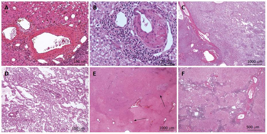
Figure 11 Inflammatory hepatocellular adenoma typical microscopic aspects.
A: Same patient as Figure 10 D. B: Same patient as Figure 10F. HE: thick-walled arteries surrounded by inflammatory cells. These pseudo portal tracts are very characteristic of inflammatory hepatocellular adenoma (IHCA). C, D: Woman born in 1972; oral contraceptives for 19 years. BMI 19.6 kg/m2. One nodule 10 cm discovered by chance. Magnetic resonance imaging: IHCA. Right hepatectomy 2009. HE: prominent sinusoidal dilatation. E, F: Same patient as Figure 10F, different tumors. E: HE - tumor ill-defined from the surrounding liver without any inflammation or sinusoidal dilatation. F: HE - the histological aspect is different with a more typical aspect of IHCA. Here, thick arteries are surrounded by inflammatory cells and fibrous tissue within the hepatocellular proliferation.
- Citation: Sempoux C, Balabaud C, Bioulac-Sage P. Pictures of focal nodular hyperplasia and hepatocellular adenomas. World J Hepatol 2014; 6(8): 580-595
- URL: https://www.wjgnet.com/1948-5182/full/v6/i8/580.htm
- DOI: https://dx.doi.org/10.4254/wjh.v6.i8.580


