Copyright
©2014 Baishideng Publishing Group Inc.
World J Hepatol. Aug 27, 2014; 6(8): 580-595
Published online Aug 27, 2014. doi: 10.4254/wjh.v6.i8.580
Published online Aug 27, 2014. doi: 10.4254/wjh.v6.i8.580
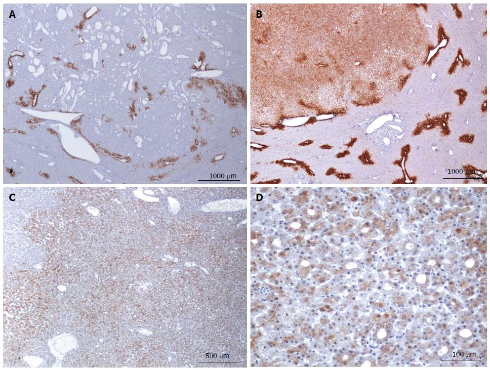
Figure 9 HNF1a inactivated hepatocellular adenoma different aspects of glutamine synthase immunostaining.
A: Same patient as Figure 7E, F. There is no staining within the lesion, except around some veins, mainly at the borders where liver-fatty acid binding protein staining (not shown) demonstrates intermingled HNF1A mutated hepatocellular adenoma (H-HCA) and normal parenchyma areas. It was impossible on HE staining to clearly identify the border of the tumor. B: Woman born in 1964; one nodule discovered by chance, 5.5 cm; oral contraceptives 21 years; BMI 18.4 kg/m2. Biopsy: β-HCA. Segmentectomy VIII 2007. Diffuse, moderate glutamine synthase (GS) staining, contrasting with normal staining in non tumoral liver in which the GS staining is limited to 1-3 rows of centrilobular hepatocytes. Today, in the absence of β-catenin nuclear staining, the significance of this abnormal GS staining in HCA remains unknown and molecular analysis is mandatory to search for β-catenin mutations. C, D: Same patient as Figure 7C, D. Heterogeneous, mild staining in tumoral hepatocytes, sometimes arranged in rosettes. The presence of rosettes is often considered as a criterion suggesting a possible malignant transformation; it may also reflect cholestatic features. Same comment as above: in the absence of molecular analysis, it is not possible to conclude and a search for β-catenin mutations is mandatory.
- Citation: Sempoux C, Balabaud C, Bioulac-Sage P. Pictures of focal nodular hyperplasia and hepatocellular adenomas. World J Hepatol 2014; 6(8): 580-595
- URL: https://www.wjgnet.com/1948-5182/full/v6/i8/580.htm
- DOI: https://dx.doi.org/10.4254/wjh.v6.i8.580


