Copyright
©2014 Baishideng Publishing Group Inc.
World J Hepatol. Aug 27, 2014; 6(8): 580-595
Published online Aug 27, 2014. doi: 10.4254/wjh.v6.i8.580
Published online Aug 27, 2014. doi: 10.4254/wjh.v6.i8.580
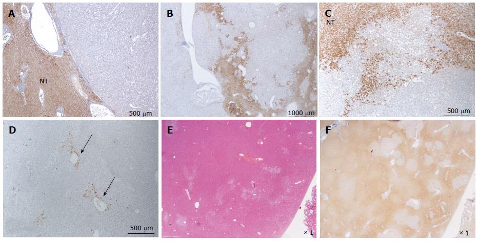
Figure 8 HNF1a inactivated hepatocellular adenoma different aspects of liver-fatty acid binding protein staining.
A: Same patient as Figure 7C, D. Typical aspect: absence of liver-fatty acid binding protein (LFABP) staining in tumor contrasting with normal expression in non tumoral liver (NT). B: Same patient as Figure 7E, F. Bands of non tumoral (NT) parenchyma normally expressing LFABP are penetrating within the unstained tumor. Indeed, the growth of HNF1A mutated hepatocellular adenoma (H-HCA) is due to the coalescence of several adenomatous liver nodules and it is not rare to find non adenomatous liver lobules squeezed in between. C: Woman born in 1962; abdominal pain. Oral contraceptives 25 years; BMI 25 kg/m2. Two liver nodules, largest 3.7 cm. Segmentectomy V/VI (2005). Bands of hepatocytes expressing LFABP within the unstained typical H-HCA. Same comment as above. D: Same patient as Figure 7E, F. LFABP expression in a few perivenular hepatocytes, particularly at the periphery of H-HCA, is a frequent observation, as well as a rim of positive hepatocytes at the border. E, F: Woman born in 1967; adenomatosis suspected during coelioscopy for extrauterine pregnancy. Segmentectomy for a 6 cm liver nodule. On HE (E), at distance from the main lesion, the presence of several steatotic small nodules well identified on LFABP staining (F). This is a very specific and characteristic aspect of H-HCA adenomatosis.s
- Citation: Sempoux C, Balabaud C, Bioulac-Sage P. Pictures of focal nodular hyperplasia and hepatocellular adenomas. World J Hepatol 2014; 6(8): 580-595
- URL: https://www.wjgnet.com/1948-5182/full/v6/i8/580.htm
- DOI: https://dx.doi.org/10.4254/wjh.v6.i8.580


