Copyright
©2014 Baishideng Publishing Group Inc.
World J Hepatol. Aug 27, 2014; 6(8): 580-595
Published online Aug 27, 2014. doi: 10.4254/wjh.v6.i8.580
Published online Aug 27, 2014. doi: 10.4254/wjh.v6.i8.580
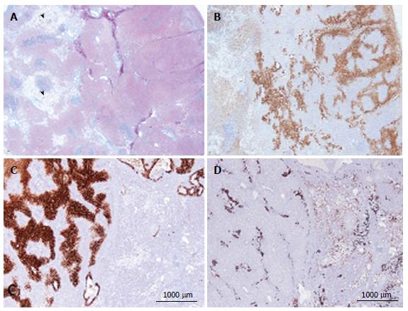
Figure 3 Focal nodular hyperplasia microscopic atypical features on Masson’s trichrome and glutamine synthase immunostaining.
A, B: Same patient as Figure 1E. A: Masson’s trichrome - large areas of sinusoidal dilatation (arrowhead), nearby solid hepatocytic areas (right) with thin, short fibrotic bands. B: Glutamine synthase (GS) immunostaining - typical aspect of focal nodular hyperplasia (FNH) with anastomosed positive areas in a “map-like pattern” in the nodular area (right); no staining in the sinusoidal dilation area (left). This aspect is very unusual. This nodule should not be interpreted as a mixed tumor (part FNH and part hepatocellular adenoma) and should not be called “telangiectatic FNH”[23,24]. A better term could be FNH with major sinusoidal dilatation. C, D: Woman born in 1969, 2003; 2 FNH: first hepatic resection (tumorectomy in 2003 for a 7-cm FNH). In 2004, persistence of abdominal discomfort (no change in size of the 7 cm FNH). Right hepatectomy. C: Typical GS staining (left); no GS staining in the area of sinusoidal dilatation (right). D: Obvious ductular reaction on the CK7 immunostaining. Although the 2 above cases are very rare, FNH with areas of sinusoidal dilatation are seen occasionally.
- Citation: Sempoux C, Balabaud C, Bioulac-Sage P. Pictures of focal nodular hyperplasia and hepatocellular adenomas. World J Hepatol 2014; 6(8): 580-595
- URL: https://www.wjgnet.com/1948-5182/full/v6/i8/580.htm
- DOI: https://dx.doi.org/10.4254/wjh.v6.i8.580


