Copyright
©2014 Baishideng Publishing Group Inc.
World J Hepatol. May 27, 2014; 6(5): 293-305
Published online May 27, 2014. doi: 10.4254/wjh.v6.i5.293
Published online May 27, 2014. doi: 10.4254/wjh.v6.i5.293
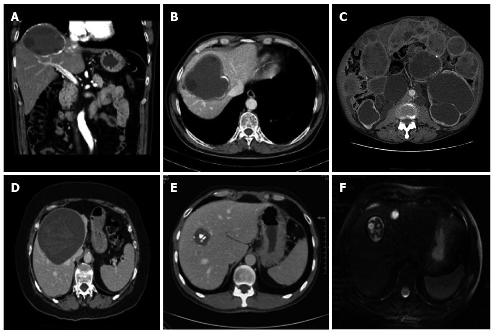
Figure 5 Computed tomography and magnetic resonance imaging of hepatic cystic echinococcosis.
A and B: Contrast enhanced computed tomography (CT) abdominal scan of a 59-year-old male patient with a CE3b cyst in the VII liver segment; C: Disseminated peritoneal echinococcosis in 64-year-old male patient, 30 years after surgery for CE without albendazole prophylaxis[1]; D: Abdominal CT scan of a 59-year-old female patient with a CE3a cyst in the IV-VIII liver segments; E: Abdominal CT scan of a 47-year-old male patient with a CE5 calcified cyst in the VIII liver segment; F: Abdominal magnetic resonance imaging scan of a 52-year-old male patient with a CE3b cyst in the VIII segment.
- Citation: Rinaldi F, Brunetti E, Neumayr A, Maestri M, Goblirsch S, Tamarozzi F. Cystic echinococcosis of the liver: A primer for hepatologists. World J Hepatol 2014; 6(5): 293-305
- URL: https://www.wjgnet.com/1948-5182/full/v6/i5/293.htm
- DOI: https://dx.doi.org/10.4254/wjh.v6.i5.293


