Copyright
©The Author(s) 2018.
World J Hepatol. Feb 27, 2018; 10(2): 186-212
Published online Feb 27, 2018. doi: 10.4254/wjh.v10.i2.186
Published online Feb 27, 2018. doi: 10.4254/wjh.v10.i2.186
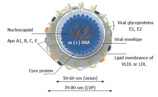
Figure 2 A model of hepatitis C virus lipoviral particle.
Lipid membrane formed by low density lipoproteins (LDL) and very low-density lipoproteins (VLDL) on the surface of the virion (given in grey). Viral core is given in blue and viral RNA is shown in orange. Heterodimers of glycoproteins E1 and E2 are partially embedded in the lipid bilayer and are forming 6 nm long spikes (projections) on the surface of the virion[14-19]. As a result of association with LDL and VLDL, the morphology of the virion is not icosahedral. Depending on the viral source, the shape and size of the particles might vary.
- Citation: Morozov VA, Lagaye S. Hepatitis C virus: Morphogenesis, infection and therapy. World J Hepatol 2018; 10(2): 186-212
- URL: https://www.wjgnet.com/1948-5182/full/v10/i2/186.htm
- DOI: https://dx.doi.org/10.4254/wjh.v10.i2.186


