Copyright
©The Author(s) 2018.
World J Hepatol. Jan 27, 2018; 10(1): 142-154
Published online Jan 27, 2018. doi: 10.4254/wjh.v10.i1.142
Published online Jan 27, 2018. doi: 10.4254/wjh.v10.i1.142
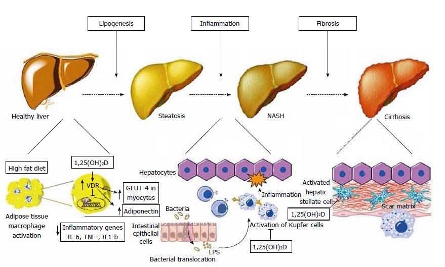
Figure 2 Schematic representation of metabolic, anti-inflammatory, and anti-fibrotic effects of vitamin D on hepatocytes and non-parenchymal hepatic cells (hepatic stellate cells, Kupffer cells) in non-alcoholic fatty liver disease.
Left: At the initial stage of lipogenesis, 1,25(OH)D acts on adipocytes and inhibits NF-κB transcription, known as the pro-inflammatory “master switch”, and thus inhibits the expression of the inflammatory cytokines IL-6, TNF-α, and IL-1β. It also increases adiponectin secretion from adipocytes and enhances GLUT-4 receptor expression in myocytes, both of which improve insulin resistance; Middle: Increased gut permeability allows the translocation of bacterial pathogens which can activate Toll-like receptors (TLR) on Kupffer cells. 1,25(OH)D downregulates the expression of TLR-2, TLR-4, and TLR-9 in these cells, thus ameliorating inflammation; Right: 1,25(OH)D acts on hepatic stellate cells by binding to VDR, which reduces the proliferation of these cells that play a major role in inducing fibrosis. VDR: Vitamin D receptor; TLR: Toll-like receptor; LPS: Lipopolysaccharide. Reproduced in compliance with Creative Commons in PubMed Central Open Access to Reproduced with the permission of the Baishideng Publishing Group Inc[9].
- Citation: Saberi B, Dadabhai AS, Nanavati J, Wang L, Shinohara RT, Mullin GE. Vitamin D levels do not predict the stage of hepatic fibrosis in patients with non-alcoholic fatty liver disease: A PRISMA compliant systematic review and meta-analysis of pooled data. World J Hepatol 2018; 10(1): 142-154
- URL: https://www.wjgnet.com/1948-5182/full/v10/i1/142.htm
- DOI: https://dx.doi.org/10.4254/wjh.v10.i1.142


