Copyright
©The Author(s) 2016.
World J Stem Cells. Dec 26, 2016; 8(12): 399-427
Published online Dec 26, 2016. doi: 10.4252/wjsc.v8.i12.399
Published online Dec 26, 2016. doi: 10.4252/wjsc.v8.i12.399
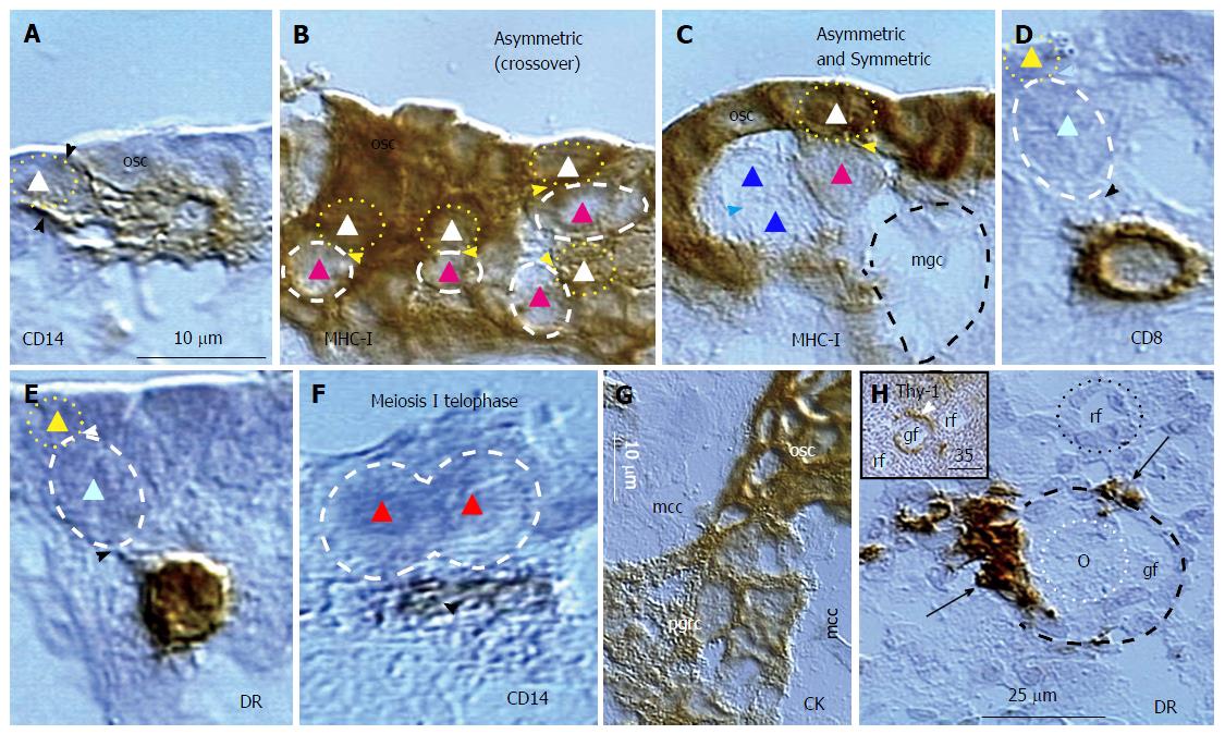
Figure 8 Origin of germ and granulosa cells in the ovary of the human midpregnancy fetus.
A: Isolated ovarian stem cell (triangle) is associated with CD14 pMDC (arrowheads); B: Fetal germ cells (red triangles) lacking MHC-I expression originate from MHC-I+ ovarian stem cells (white triangles); C: The symmetric division of germ cells (blue triangles) follows asymmetric division (white and red triangles) of ovarian stem cells, and causes emergence of moving germ cell (mgc); D: The origin of germ cells requires association of CD8+ (D) and DR+ (E) T cell; F: Meiosis I telophase (red triangles) is associated with a primitive MDC (arrowhead); G: The fetal primitive granulosa cells (pgrc) originate from ovarian stem cells between the mesenchymal cell cords (mcc); H: Small growing follicle (gf) is accompanied by DR+ MDCs (arrows), which are absent at the resting follicle (rf). Inset shows Thy-1+ pericytes (arrowhead) during follicular selection (Figure 7). Bar in A for A-F. Adjusted from[44] with a permission: ©Springer United States. MDC: Monocyte-derived cell; pMDC: Primitive MDC.
- Citation: Bukovsky A. Involvement of blood mononuclear cells in the infertility, age-associated diseases and cancer treatment. World J Stem Cells 2016; 8(12): 399-427
- URL: https://www.wjgnet.com/1948-0210/full/v8/i12/399.htm
- DOI: https://dx.doi.org/10.4252/wjsc.v8.i12.399


