Copyright
©The Author(s) 2016.
World J Stem Cells. Dec 26, 2016; 8(12): 399-427
Published online Dec 26, 2016. doi: 10.4252/wjsc.v8.i12.399
Published online Dec 26, 2016. doi: 10.4252/wjsc.v8.i12.399
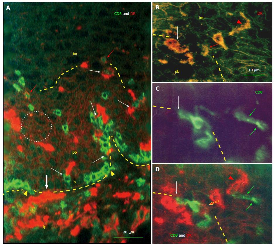
Figure 3 Interaction of monocyte derived cells and T cells in the squamous epithelium.
A: HLA DR+ monocyte-derived cells (MDCs) (red color) and CD8 T cells (TC) (green color) enter (bold white arrow and arrowheads) basal epithelial layer (b) from the lamina propria (lp). Note accumulation of TC among basal epithelial cells. Both cell types interact by themselves (white arrows). After reaching the parabasal (pb)/intermediate (im) interface (dashed line) TC also express DR (red arrows) indicating their activation. Note binding of DR released from MDCs (see Figure 2A) to parabasal epithelial cells (doted circle); B-D: Detail of pb/im interface shows MDC/T cell interaction (white arrows), the DR expression by TC (red arrows), transition of MDC into elongated dendritic cell (arrowheads), and remnants of regressing T cell in the lower intermediate layer (green arrows)[14].
- Citation: Bukovsky A. Involvement of blood mononuclear cells in the infertility, age-associated diseases and cancer treatment. World J Stem Cells 2016; 8(12): 399-427
- URL: https://www.wjgnet.com/1948-0210/full/v8/i12/399.htm
- DOI: https://dx.doi.org/10.4252/wjsc.v8.i12.399


